


1. CELL AND CELL THEORY
- Biology is the study of living organisms. It is the cell theory that emphasised the unity underlying this diversity of forms, i.e., the cellular organisation of all life forms. Cell theory also created a sense of mystery around living phenomena, i.e., physiological and behavioural processes. This mystery was the requirement of integrity of cellular organisation for living phenomena to be demonstrated or observed. This approach enables us to describe the various processes in molecular terms. The approach is established by analysis of living tissues for elements and compounds. It will tell us what types of organic compounds are present in living organisms.
- This physico-chemical approach to study and understand living organisms is called ‘Reductionist Biology’. The concepts and techniques of physics and chemistry are applied to understand biology.
- N. RAMACHANDRAN, an outstanding figure in the field of protein structure, was the founder of the ‘Madras school’ of conformational analysis of biopolymers.
- His discovery of the triple helical structure of collagen published in Nature in 1954 and his analysis of the allowed conformations of proteins through the use of the ‘Ramachandran plot’ rank among the most outstanding contributions in structural biology.
- Ramachandran met Linus Pauling and was deeply influenced by his publications on models of the α-helix and β-sheet structures that directed his attention to solving the structure of collagen.
- When you look around, you see both living and non-living things. You must have wondered and asked yourself – ‘what is it that makes an organism living, or what is it that an inanimate thing does not have which a living thing has’ ? The answer to this is the presence of the basic unit of life – the cell in all living organisms. All organisms are composed of cells. Some are composed of a single cell and are called unicellular organisms while others, like us, composed of many cells, are called multicellular organisms.
What is a cell?
- Unicellular organisms are capable of (i) independent existence and (ii) performing the essential functions of life. Anything less than a complete structure of a cell does not ensure independent living. Hence, cell is the fundamental structural and functional unit of all living organisms. Anton Von Leeuwenhoek first saw and described a live cell. Robert Brown later discovered the nucleus. The invention of the microscope and its improvement leading to the electron microscope revealed all the structural details of the cell.
Cell Theory
- In 1838, Malthias Schleiden, a German botanist, examined a large number of plants and observed that all plants are composed of different kinds of cells which form the tissues of the plant.
- At about the same time, Theodore Schwann (1839), a British Zoologist, studied different types of animal cells and reported that cells had a thin outer layer which is today known as the ‘plasma membrane’.
- He also concluded, based on his studies on plant tissues, that the presence of cell wall is a unique character of the plant cells. On the basis of this, Schwann proposed the hypothesis that the bodies of animals and plants are composed of cells and products of cells.
- Schleiden and Schwann together formulated the cell theory. This theory however, did not explain as to how new cells were formed.
- Rudolf Virchow (1855) first explained that cells divided and new cells are formed from pre-existing cells (Omnis cellula-e cellula). He modified the hypothesis of Schleiden and Schwann to give the cell theory a final shape. Cell theory as understood today is:
(i) all living organisms are composed of cells and products of cells.
(ii) all cells arise from pre-existing cells.
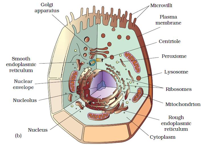
An Overview of Cell
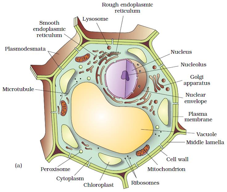
- The onion cell which is a typical plant cell, has a distinct cell wall as its outer boundary and just within it is the cell membrane. The cells of the human cheek have an outer membrane as the delimiting structure of the cell.
- Inside each cell is a dense membrane bound structure called nucleus. This nucleus contains the chromosomes which in turn contain the genetic material, DNA.
- Cells that have membrane bound nuclei are called eukaryotic whereas cells that lack a membrane bound nucleus are prokaryotic.
- In both prokaryotic and eukaryotic cells, a semi-fluid matrix called cytoplasm occupies the volume of the cell. The cytoplasm is the main arena of cellular activities in both the plant and animal cells. Various chemical reactions occur in it to keep the cell in the ‘living state’.
- Besides the nucleus, the eukaryotic cells have other membrane bound distinct structures called organelles like the endoplasmic reticulum (ER), the Golgi complex, lysosomes, mitochondria, microbodies and vacuoles.
- The prokaryotic cells lack such membrane bound organelles. Ribosomes are non-membrane bound organelles found in all cells – both eukaryotic as well as prokaryotic. Within the cell, ribosomes are found not only in the cytoplasm but also within the two organelles – chloroplasts (in plants) and mitochondria and on rough ER.
- Animal cells contain another non-membrane bound organelle called centriole which helps in cell division.
- Cells differ greatly in size, shape and activities. For example, Mycoplasmas, the smallest cells, are only 0.3 μm in length while bacteria could be 3 to 5 μm. The largest isolated single cell is the egg of an ostrich.
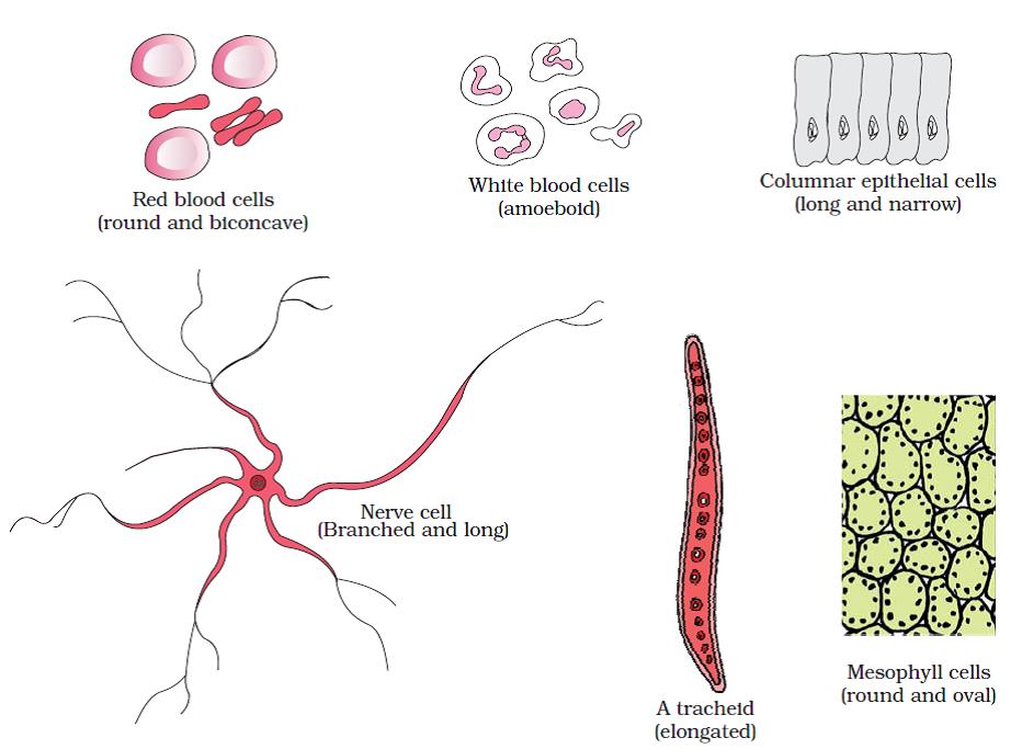
Diagram showing different shapes of the cells - Among multicellular organisms, human red blood cells are about 7.0 μm in diameter. Nerve cells are some of the longest cells. Cells also vary greatly in their shape. They may be disc-like, polygonal, columnar, cuboid, thread like, or even irregular. The shape of the cell may vary with the function they perform.
2. PROKARYOTIC CELLS
- The prokaryotic cells are represented by bacteria, blue-green algae, mycoplasma and PPLO (Pleuro Pneumonia like Organisms). They are generally smaller and multiply more rapidly than the eukaryotic cells. They may vary greatly in shape and size. The four basic shapes of bacteria are bacillus (rod like), coccus (spherical), vibrio (comma shaped) and spirillum (spiral).
- The organisation of the prokaryotic cell is fundamentally similar even though prokaryotes exhibit a wide variety of shapes and functions.
- All prokaryotes have a cell wall surrounding the cell membrane. The fluid matrix filling the cell is the cytoplasm. There is no well-defined nucleus. The genetic material is basically naked, not enveloped by a nuclear membrane.
- In addition to the genomic DNA (the single chromosome/circular DNA), many bacteria have small circular DNA outside the genomic DNA. These smaller DNA are called plasmids. The plasmid DNA confers certain unique phenotypic characters to such bacteria. One such character is resistance to antibiotics. This plasmid DNA is used to monitor bacterial transformation with foreign DNA.
- No organelles, like the ones in eukaryotes, are found in prokaryotic cells except for ribosomes. Prokaryotes have something unique in the form of inclusions.
- A specialised differentiated form of cell membrane called mesosome is the characteristic of prokaryotes. They are essentially infoldings of cell membrane.
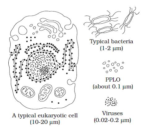
Diagram showing comparison of eukaryotic cell with other organisms
Cell Envelope and Its Modifications
- Most prokaryotic cells, particularly the bacterial cells, have a chemically complex cell envelope. The cell envelope consists of a tightly bound three layered structure i.e., the outermost glycocalyx followed by the cell wall and then the plasma membrane. Although each layer of the envelope performs distinct function, they act together as a single protective unit.
- Bacteria can be classified into two groups on the basis of the differences in the cell envelopes and the manner in which they respond to the staining procedure developed by Gram viz., those that take up the gram stain are Gram positive and the others that do not are called Gram negative
- Glycocalyx differs in composition and thickness among different bacteria. It could be a loose sheath called the slime layer in some, while in others it may be thick and tough, called the capsule.
- The cell wall determines the shape of the cell and provides a strong structural support to prevent the bacterium from bursting or collapsing.
- The plasma membrane is semi-permeable in nature and interacts with the outside world. This membrane is similar structurally to that of the eukaryotes.
- A special membranous structure is the mesosome which is formed by the extensions of plasma membrane into the cell. These extensions are in the form of vesicles, tubules and lamellae. They help in cell wall formation, DNA replication and distribution to daughter cells. They also help in respiration, secretion processes, to increase the surface area of the plasma membrane and enzymatic content.
- In some prokaryotes like cyanobacteria, there are other membranous extensions into the cytoplasm called chromatophores which contain pigments.
- Bacterial cells may be motile or non-motile. If motile, they have thin filamentous extensions from their cell wall called flagella. Bacteria show a range in the number and arrangement of flagella. Bacterial flagellum is composed of three parts – filament, hook and basal body. The filament is the longest portion and extends from the cell surface to the outside.
- Besides flagella, Pili and Fimbriae are also surface structures of the bacteria but do not play a role in motility. The pili are elongated tubular structures made of a special protein. The fimbriae are small bristle like fibres sprouting out of the cell. In some bacteria, they are known to help attach the bacteria to rocks in streams and also to the host tissues.
Ribosomes and Inclusion Bodies
- In prokaryotes ribosomes are associated with the plasma membrane of the cell. They are about 15 nm by 20 nm in size and are made of two subunits – 50S and 30S units which when present together form 70S prokaryotic ribosomes. Ribosomes are the site of protein synthesis. Several ribosomes may attach to a single mRNA and form a chain called polyribosomes or polysome. The ribosomes of a polysome translate the mRNA into proteins.
- Inclusion bodies: Reserve material in prokaryotic cells are stored in the cytoplasm in the form of inclusion bodies. These are not bounded by any membrane system and lie free in the cytoplasm, e.g., phosphate granules, cyanophycean granules and glycogen granules. Gas vacuoles are found in blue green and purple and green photosynthetic bacteria.
3. EUKARYOTIC CELLS
- The eukaryotes include all the protists, plants, animals and fungi. In eukaryotic cells there is an extensive compartmentalisation of cytoplasm through the presence of membrane bound organelles.
- Eukaryotic cells possess an organised nucleus with a nuclear envelope. In addition, eukaryotic cells have a variety of complex locomotory and cytoskeletal structures.
- Their genetic material is organised into chromosomes. All eukaryotic cells are not identical. Plant and animal cells are different as the former possess cell walls, plastids and a large central vacuole which are absent in animal cells. On the other hand, animal cells have centrioles which are absent in almost all plant cells.
4. CELL MEMBRANE
- Its presence was first recognised by Nageli & Cramer (1855) who gave the term ‘plasma membrane’
- It was experimentally confirmed by Overton (1899) suggesting that it is made of lipid.
- O. Plowe (1931) called it plasmalemma.
- Gorter and Grendel (1925) postulated that it is made of “bimolecular lipid layer”.
- Every cell is bounded by membranous covering which separates, the contents of cells from their external environment, controlling exchange of materials such as nutrients and waste products between the two. They also enable separate compartments to be formed inside cells in which specialised metabolic processes such as photosynthesis and aerobic respiration can take place.
- Membranes also act as receptor sites for recognising hormones, neurotransmitters and other chemicals, either from the external environment or from other parts of the organism.
- A thin, delicate membrane of about 70 Å to 100 Å thickness made almost entirely of proteins and lipids. The most common lipids are phospholipids.
- Each phospholipid molecule consists of a polar head containing phosphate, and two non-polar hydrocarbon tails from the fatty acids used to make the molecule. The head is hydrophilic (water-loving) and the tails are hydrophobic (water-hating). In presence of water they form a bilayer.
- Membrane proteins have been classified as integral (intrinsic) or peripheral (extrinsic) according to the degree of their association with the membrane and the methods by which they can be solubilized.
- Peripheral proteins are separated by mild treatment, are soluble in aqueous solutions, and are usually free of lipids e.g. spectrin of erythrocytes, cytochrome c of mitochondria.
- Integral proteins represents more than 70% of the membrane proteins and require drastic procedures for isolation. e.g. most membrane-bound enzymes, drug and hormone receptors, histocompatibility antigens (glycophorins).
- Various models about its structure are based upon its biochemical and biophysical properties:
- Lamellar or Sandwitch Model : by Danielli and Davson According to this two layers of phospholipids were covered with proteins on the two sides. Phospholipid molecules are at right angles to the plane of the membrane. Their charged hydrophilic heads are associated with the ionized groups of the protein, present in the form of thin sheets. The sheets of protein and hydrophobic properties of the membrane make it selectively permeable.
- Unit Membrane Concept : Robertson (1959) found in his EM studies, membrane as a three layers (trilaminar) structure consisting of two electron-dense zones of 2 nm thickness, separated by a middle light zone of 3.5 nm width. He also supported the model of Danielli and Davson with little modification.
- The dark zone consists of protein and head part of lipid and middle light zone consists of the tail part of lipid of both the layers together. Also called as “rail-track”
- The three-layered-protein membrane is a fundamental structural unit in all the cells, termed as ‘unit membrane’ to denote its cellular universality.
- The cell membrane consists of a single unit membrane while the double membrane coverings of the mitochondrion, chloroplast, endoplasmic reticulum, nucleus and Golgi apparatus consists of two unit membranes in paired arrangement and in close apposition. The grana of the chloroplast is interpreted as multilayered structure made up of unit membranes closely packed and stacked on top of another.
- The detailed structure of the membrane was studied only after the advent of the electron microscope in the 1950s.
- Meanwhile, chemical studies on the cell membrane, especially in human red blood cells (RBCs), enabled the scientists to deduce the possible structure of plasma membrane. These studies showed that the cell membrane is composed of lipids that are arranged in a bilayer. Also, the lipids are arranged within the membrane with the polar head towards the outer sides and the hydrophobic tails towards the inner part. This ensures that the nonpolar tail of saturated hydrocarbons is protected from the aqueous environment.
- The lipid component of the membrane mainly consists of phosphoglycerides.
- Later, biochemical investigation clearly revealed that the cell membranes also possess protein and carbohydrate.
- The ratio of protein and lipid varies considerably in different cell types. In human beings, the membrane of the erythrocyte has approximately 52 per cent protein and 40 per cent lipids.
- Depending on the ease of extraction, membrane proteins can be classified as integral or peripheral. Peripheral proteins lie on the surface of membrane while the integral proteins are partially or totally buried in the membrane.
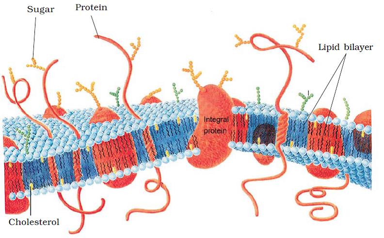
Fluid mosaic model of plasma membran - An improved model of the structure of cell membrane was proposed by Singer and Nicolson (1972) widely accepted as fluid mosaic model. According to this, the quasi-fluid nature of lipid enables lateral movement of proteins within the overall bilayer. This ability to move within the membrane is measured as its fluidity. The fluid nature of the membrane is also important from the point of view of functions like cell growth, formation of intercellular junctions, secretion, endocytosis, cell division etc.
- One of the most important functions of the plasma membrane is the transport of the molecules across it. The membrane is selectively permeable to some molecules present on either side of it.
- Many molecules can move briefly across the membrane without any requirement of energy and this is called the passive transport.
- Neutral solutes may move across the membrane by the process of simple diffusion along the concentration gradient, i.e., from higher concentration to the lower. Water may also move across this membrane from higher to lower concentration. Movement of water by diffusion is called osmosis.
- As the polar molecules cannot pass through the nonpolar lipid bilayer, they require a carrier protein of the membrane to facilitate their transport across the membrane. A few ions or molecules are transported across the membrane against their concentration gradient, i.e., from lower to the higher concentration. Such a transport is an energy dependent process, in which ATP is utilised and is called active transport, e.g., Na+/K+
Transport across the cell surface membrane
- Cell membranes are only about 7 nm wide but they present barriers to the movement of ions and molecules, particularly polar (water-soluble) molecules such as glucose and amino acids that are repelled by the non-polar, hydrophobic lipids of membranes. This prevents the aqueous contents of the cell from escaping.
- Transport across membranes must still occur to
- to obtain nutrients.
- to excrete waste substances.
- to secrete useful substances.
- to generate the ionic gradients essential for nervous and muscular activity.
- to maintain a suitable pH and ionic concentration within the cell for enzyme activity.
There are four basic mechanisms, namely diffusion, osmosis, active transport and bulk transport.
Diffusion
- Movement of molecules or ions from a region of their higher concentration to a region of their lower concentration down a diffusion gradient. The process is passive; it does not require energy and happens spontaneously
- Occurs by the random motion of molecules which is due to their kinetic energy (energy of movement). Each type of molecule moves down its own diffusion gradient independently of other molecules. e.g. oxygen diffuses from the lungs into the blood while at the same time carbon dioxide diffuses in the opposite direction.
Three factors in particular affect the rate of diffusion:
1. The steepness of the diffusion gradient, the steeper the gradient, the faster the rate of diffusion.
2. The greater the surface area of a membrane through which diffusion is taking place, the greater the rate of diffusion.
3. Rate of diffusion decreases rapidly with distance. Diffusion is therefore only effective over very short distances. It is essential that membranes are thin so that molecules or ions can cross them rapidly. - The respiratory gases, oxygen and carbon dioxide, diffuse rapidly through membranes. Water molecules, although very polar, are small enough to pass between the hydrophobic phospholipid molecules without interference. However, ions and larger polar molecules such as amino acids, sugars, fatty acids and glycerol are repelled by the hydrophobic region of the membrane and diffuse across extremely slowly.
Facilitated Diffusion
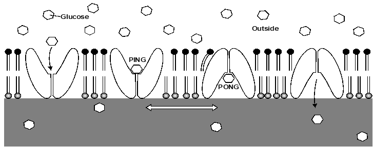
Facilitated diffusion through a carrier protein
- Some ions and polar molecules can diffuse through special transport proteins called channel proteins and carrier proteins.
- Channel proteins contain water-filled hydrophilic channels or pores whose shape is specific for a particular ion or molecule. Alternately several proteins combine, forming a channel between them.
- Channel proteins have a fixed shape. Diffusion can occur through the channel in either direction.
- Disease cystic fibrosis is caused by a fault in a protein which acts as a chloride ion channel.
- Carrier proteins undergo rapid changes in shape. They exist in two forms, known as the ‘ping’ and ‘pong’ states. The binding site faces outwards in one state and into the cell in the other state.
Osmosis
- The passage of water molecules from a region of their higher concentration to a region of their lower concentration through a partially permeable membrane. A form of diffusion in which only water molecules move.
- The tendency of water molecules to move from one place to another is measured as the water potential, represented by the symbol (Greek letter psi). Water always moves from a region of higher water potential to one of lower water potential. Solute molecules reduce. The extent by which they lower is known as the solute potential.
Active Transport
- Active transport is the energy-consuming transport of molecules or ions across a membrane against a concentration gradient.
- Energy is required because the substance must be moved against its natural tendency to diffuse in the opposite direction.
- Movement is usually in one direction only, unlike diffusion which is reversible.
- Active transport is achieved by carrier proteins situated in the cell surface membrane.
- The carrier proteins involved in active transport need a supply of energy to keep changing shape which is provided by ATP from respiration.
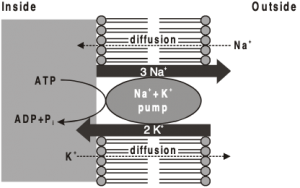
Sodium Potassium Pump (Na+ – K+ Pump)
- The pump is a carrier protein which spans the membrane from one side to the other.
- On the inside it accepts sodium and ATP, while on the outside it accepts potassium. For every 2K+ taken into the cell, 3Na+ are removed.
- The transfer of sodium and potassium across the membrane is brought about by changes in the shape of the protein.
- A potential difference is built up across the membrane, with the inside of the cell being negative.
- Potential difference tends to restrict the entry of negatively charged ions (anions) such as chloride which explains why the chloride concentration inside red blood cells is less than outside despite the fact that chloride can diffuse in and out by facilitated diffusion.
- Positively charged ions (cations) tend to be attracted into cells.
- Both concentration and charge are important in deciding the direction in which ions cross membranes.
- Essential in controlling the osmotic balance of animal cells (osmoregulation).
- If the pump is inhibited, the cell swells and bursts because a build-up of sodium ions results in excess water entering the cells by osmosis.
- Important in maintaining electrical activity in nerve and muscle cells and in driving active transport of some other substances such as sugars and amino acids.
- Also, high concentrations of potassium are needed inside cells for protein synthesis, glycolysis, photosynthesis and other vital processes.
- Active transport is associated with epithelial cells, as in the gut lining and kidney tubules (nephrons), because these are active in secretion and absorption.
Bulk Transport
Endocytosis & Exocytosis
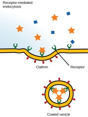
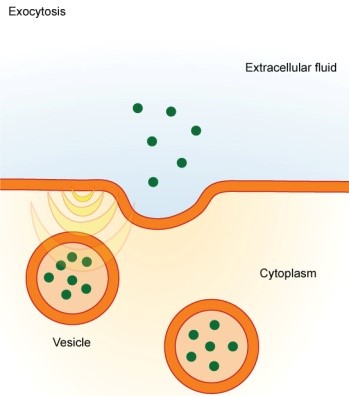
- Endocytosis and exocytosis are active processes involving the bulk transport of materials through membranes, either into cells (endocytosis) or out of cells (exocytosis).
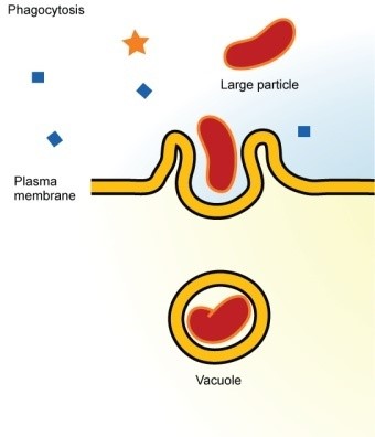
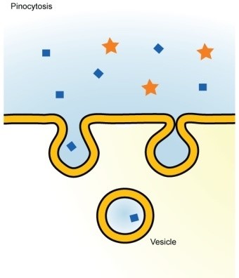
- Endocytosis occurs by an infolding or extension of the cell surface membrane to form a vesicle or vacuole. It is of two types:
1. Phagocytosis (‘cell eating’) Material taken up is in solid form. Cells specialising in the process are called phagocytes and are said to be phagocytic. e.g., some white blood cells take up bacteria by phagocytosis. The sac formed during uptake is called a phagocytic vacuole.
2. Pinocytosis (‘cell drinking’) Material taken up is in liquid form. Vesicles formed are often extremely small. Pinocytosis is used by the human egg cell to take up nutrients from the surrounding follicle cells.
Exocytosis is the reverse process of endocytosis. Waste materials may be removed from cells, such as solid, undigested remains from phagocytic vacuoles, or useful materials may be secreted. Secretion of enzymes from the pancreas is achieved in this way. Plant cells use exocytosis to export the materials needed to form cell walls.
 |
 |
 |
 |
5. CELL WALL
- A non-living rigid structure called the cell wall forms an outer covering for the plasma membrane of fungi and plants. Cell wall not only gives shape to the cell and protects the cell from mechanical damage and infection, it also helps in cell-to-cell interaction and provides barrier to undesirable macromolecules.
- Algae have cell wall, made of cellulose, galactans, mannans and minerals like calcium carbonate, while in other plants it consists of cellulose, hemicellulose, pectins and proteins.
- The cell wall of a young plant cell, the primary wall is capable of growth, which gradually diminishes as the cell matures and the secondary wall is formed on the inner (towards membrane) side of the cell.
- The middle lamella is a layer mainly of calcium pectate which holds or glues the different neighbouring cells together. The cell wall and middle lamellae may be traversed by plasmodesmata which connect the cytoplasm of neighbouring cells.
Composition
- Consist of a microfibrillar network lying in a gel-like matrix of interlinked molecules.
- The microfibrils are mostly cellulose chains which by intra and intermolecular hydrogen bonding produce the structural unit known as microfibril.
- Each microfibril is 25 nm in diameter and is composed of about 2000 glucan chains. Microfibrils in turn associate among themselves and with non-cellulosic polysaccharides and proteins to form the cell wall.
- Gel matrix, in addition to protein, contains some polysaccharides (Pectin substances and hemicelluloses) and lignin (found only in mature cell walls).
- Certain cell walls may have cuticular substance (cutin wax) and mineral deposits in the form of calcium and magnesium carbonates and silicates.
- In fungi and yeasts the cell wall is composed of chitin, a polymer of N-acetyl glucosamine.
Formation
Begins at late anaphase and continues through the telophase.
- The cell plate which is formed from vesicles of the Golgi complex becomes aligned at the equatorial plane and forms the intercellular layer or middle lamella.
- After cell division, each daughter cell deposits other layer that constitutes the primary cell wall, when the cell has enlarged to its mature size usually with the development of large vacuoles, the secondary wall appears by apposition of new materials on the primary cell wall.
Structure
The cell wall is differentiated into three layers :
(a) Middle lamella : Binds the two adjacent cells firmly.
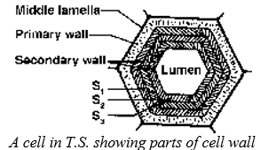
It is an amorphous layer composed of calcium and magnesium pectate. In the softer tissues of the plant e.g. pith, cambium, etc., it remains unlignified while in woody tissues it is highly lignified. In fleshy fruits during ripening the pectin of the middle lamella is dissolved by the proteolytic enzymes, the cells are, therefore, loosened and the fruit becomes soft.
(b) Primary wall : Primary wall is thin and elastic capable of growth and extension, composed largely of cellulose with some pectic substances. Becomes lignified and thick after growth stops.
(c) Secondary wall : Secondary wall particularly of sclerenchyma is very thick and rigid and consists of three layers — outer (S1), middle (S2) and inner (S3) layers. The cellulose microfibrils of these layers are embedded in a matrix composed of hemicellulose, lignin, pectin etc.
Sometimes secondary wall layers are not deposited in the adjacent cells and such thin spots are called simple pits. The middle lamella and the two primary walls in the pit region together constitute the pit membrane, which has a thickening called torus.
ENDOMEMBRANE SYSTEM
While each of the membranous organelles is distinct in terms of its structure and function, many of these are considered together as an endomembrane system because their functions are coordinated. The endomembrane system include endoplasmic reticulum (ER), Golgi complex, lysosomes and vacuoles. Since the functions of the mitochondria, chloroplast and peroxisomes are not coordinated with the above components, these are not considered as part of the endomembrane system.
6. THE ENDOPLASMIC RETICULUM (ER)
- Electron microscopic studies of eukaryotic cells reveal the presence of a network or reticulum of tiny tubular structures scattered in the cytoplasm that is called the endoplasmic reticulum (ER). Hence, ER divides the intracellular space into two distinct compartments, i.e., luminal (inside ER) and extra luminal (cytoplasm) compartments.
- The ER often shows ribosomes attached to their outer surface. The endoplasmic reticulum bearing ribosomes on their surface is called rough endoplasmic reticulum (RER). In the absence of ribosomes they appear smooth and are called smooth endoplasmic reticulum (SER).
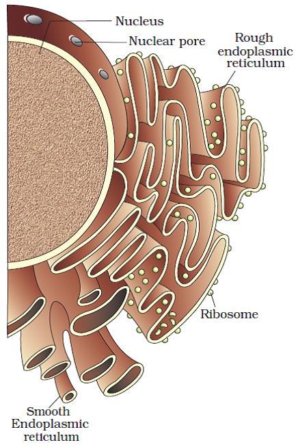
- RER is frequently observed in the cells actively involved in protein synthesis and secretion. They are extensive and continuous with the outer membrane of the nucleus.
- The smooth endoplasmic reticulum is the major site for synthesis of lipid. In animal cells lipid-like steroidal hormones are synthesised in SER.
Function of SER
- Intracellular transport of substances.
- Storage of glycogen, lipid, calcium.
- Glycosidation or synthesis of glycoprotein, glycolipids etc.
- Detoxification of various toxic, waste materials like drugs, pollutants, bile salts, degraded amino acids and fatty acids.
- Provides skeletal framework to the cell and gives rise to membrane organelles like Golgi bodies, spherosomes.
- Plays important role in cycling of membranes.
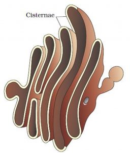
7. GOLGI APPARATUS
- Camillo Golgi (1898) first observed densely stained reticular structures near the nucleus. These were later named Golgi bodies after him.
- They consist of many flat, disc-shaped sacs or cisternae of 0.5μm to 1.0μm diameter. These are stacked parallel to each other. Varied number of cisternae are present in a Golgi complex.
- The Golgi cisternae are concentrically arranged near the nucleus with distinct convex cis or the forming face and concave trans or the maturing face.
|
|
|
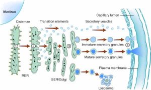
The cis and the trans faces of the organelle are entirely different, but interconnected.
- The Golgi apparatus principally performs the function of packaging materials, to be delivered either to the intra-cellular targets or secreted outside the cell.
- Materials to be packaged in the form of vesicles from the ER fuse with the cis face of the Golgi apparatus and move towards the maturing face. This explains why the Golgi apparatus remains in close association with the endoplasmic reticulum.
- A number of proteins synthesised by ribosomes on the endoplasmic reticulum are modified in the cisternae of the Golgi apparatus before they are released from its trans Golgi apparatus is the important site of formation of glycoproteins and glycolipids.
- Formation of primary lysosome.
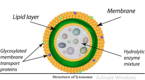
8. LYSOSOMES
- These are membrane bound vesicular structures formed by the process of packaging in the Golgi apparatus.
- The isolated lysosomal vesicles have been found to be very rich in almost all types of hydrolytic enzymes (hydrolases – lipases, proteases, carbohydrases) optimally active at the acidic pH. These enzymes are capable of digesting carbohydrates, proteins, lipids and nucleic acids.
- Discovered by Christian de Duve (1955) from liver cells of rats.
- Forms GERL complex with Golgibodies and ER.
- Popularly called as “suicide bags”, occur in all the animal cells (except R.B.C.), Euglena, yeast and fungi.
Types
- Primary lysosomes : Arise from Golgi bodies, contain enzymes synthesised in the RER.
- Secondary lysosomes : Known as digestive vacuoles after primary lysosomes combine with phagosome.
- Autophagic vacuoles : During starvation the primary lysosomes become autophagic because they engulf and digest the cell organelles.
- Residual bodies : Secondary lysosomes as well as the autophagic vacuoles containing undigested matter.
Functions
- Primarily meant for heterotrophic nutrition.
- Being abundant in leucocytes, these help in defence against microbes etc.
- Help in fertilization, differentiation and metamorphosis by performing histolysis.
- Dispose off the worn out, damaged and dead cells.
- In osteoclasts, help in replacing cartilage and unwanted part of bone.
- There are a variety of lysosomal deficiency diseases; due to lack of certain enzymes storage of various unwanted lipids and carbohydrates occurs in cells causing deleterious metabolic errors.
9. VACUOLES
- The vacuole is the membrane-bound space found in the cytoplasm.
- It contains water, sap, excretory product and other materials not useful for the cell.
- The vacuole is bound by a single membrane called tonoplast.
- In plant cells the vacuoles can occupy up to 90 per cent of the volume of the cell. In plants, the tonoplast facilitates the transport of a number of ions and other materials against concentration gradients into the vacuole, hence their concentration is significantly higher in the vacuole than in the cytoplasm.
- In Amoeba the contractile vacuole is important for excretion. In many cells, as in protists, food vacuoles are formed by engulfing the food particles.
10. MITOCHONDRIA
- Mitochondria (sing.: mitochondrion), unless specifically stained, are not easily visible under the microscope. The number of mitochondria per cell is variable depending on the physiological activity of the cells.
- In terms of shape and size also, considerable degree of variability is observed. Typically it is sausage-shaped or cylindrical having a diameter of 0.2-1.0μm (average 0.5μm) and length 1.0-4.1μm.
- Each mitochondrion is a double membrane-bound structure with the outer membrane and the inner
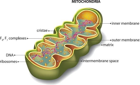
membrane dividing its lumen distinctly into two aqueous compartments, i.e., the outer compartment and the inner compartment. The inner compartment is called the matrix. The outer membrane forms the continuous limiting boundary of the organelle.
- The inner membrane forms a number of infoldings called the cristae (sing.: crista) towards the matrix. The cristae increase the surface area. The two membranes have their own specific enzymes associated with the mitochondrial function. Mitochondria are the sites of aerobic respiration. They produce cellular energy in the form of ATP, hence they are called ‘power houses’ of the cell.
- The matrix also possesses single circular DNA molecule, a few RNA molecules, ribosomes (70S) and the components required for the synthesis of proteins. The mitochondria divide by fission.
- Mitochondria have character of changing structure. This is dependent upon the physiological activity of the cell as well as the activity occurring in the organelle.
- Inactive or orthodox state, when ATP concentration is low or the respiratory chain is inhibited, the matrix of the mitochondria occupies a larger area.
- Active or condensed state when mitochondria are actively engaged in photo-phosphorylation and electron transport, the cristae are more randomly distributed and the inter-membrane space remains highly enlarged.
Functions
- Known as the ‘power house of cell’, the energy is released in it through the cellular respiration, present in the aerobic organisms only.
- Within the cell, the site of respiration or oxidative phosphorylation is mitochondria.
- The end product of glycolysis is pyruvic acid which enters mitochondria and takes part in Krebs’ cycle in the mitochondrial matrix.
11. PLASTIDS
- Plastids are found in all plant cells and in euglenoides. These are easily observed under the microscope as they are large. They bear some specific pigments, thus imparting specific colours to the plants.
- Based on the type of pigments plastids can be classified into chloroplasts, chromoplasts and
- The chloroplasts contain chlorophyll and carotenoid pigments which are responsible for trapping light energy essential for photosynthesis.
- In the chromoplasts fat soluble carotenoid pigments like carotene, xanthophylls and others are present. This gives the part of the plant a yellow, orange or red colour.
- The leucoplasts are the colourless plastids of varied shapes and sizes with stored nutrients: Amyloplasts store carbohydrates (starch), e.g., potato; elaioplasts store oils and fats whereas the aleuroplasts store proteins.
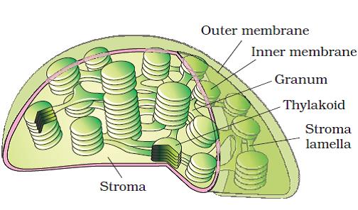
- Majority of the chloroplasts of the green plants are found in the mesophyll cells of the leaves. These are lens-shaped, oval, spherical, discoid or even ribbon-like organelles having variable length (5-10 mm) and width (2-4mm).
- Their number varies from 1 per cell of the Chlamydomonas, a green alga to 20-40 per cell in the mesophyll.
- Like mitochondria, the chloroplasts are also double membrane bound. Of the two, the inner chloroplast membrane is relatively less permeable. The space limited by the inner membrane of the chloroplast is called the stroma. A number of organised flattened membranous sacs called the thylakoids, are present in the stroma. Thylakoids are arranged in stacks like the piles of coins called grana (singular: granum) or the intergranal thylakoids. In addition, there are flat membranous tubules called the stroma lamellae connecting the thylakoids of the different grana. The membrane of the thylakoids enclose a space called a lumen.
- The stroma of the chloroplast contains enzymes required for the synthesis of carbohydrates and proteins. It also contains small, double-stranded circular DNA molecules and ribosomes. Chlorophyll pigments are present in the thylakoids. The ribosomes of the chloroplasts are smaller (70S) than the cytoplasmic ribosomes (80S).
Similarities Between Mitochondria And Chloroplasts
(i) Mitochondria and Chloroplasts may have existed as independent organisms, which during evolution developed a symbiotic relationship with plant and animal cells and evolved into their present state.
(ii) Both originate and develop in the same way and are formed by the division of pre-existing organelles.
(iii) Both are semi-autonomus organelles as both contain DNA, RNA and ribosomes. Both mitochondrial DNA (mt DNA) and chloroplast DNA (cpDNA), are circular in shape although cpDNA is much bigger than mtDNA. The genetic information contained in mtDNA and cpDNA is limited.
(iv) The symbiont hypothesis suggests that there are many similarities between prokaryotes on one hand, and mitochondria and chloroplasts on the other, e.g. the presence of circular DNA not associated with histones and 70 S ribosomes.
12. RIBOSOMES
Ribosomes are the granular structures first observed under the electron microscope as dense particles by George Palade (1953). They are composed of ribonucleic acid (RNA) and proteins and are not surrounded by any membrane. The eukaryotic ribosomes are 80S while the prokaryotic ribosomes are 70S. Here ‘S’ stands for the sedimentation coefficient; it indirectly is a measure of density and size. Both 70S and 80S ribosomes are composed of two subunits.
- Two subunits are held together by Mg+2 which form ionic bond with phosphate groups of r-RNA of two subunits. Minimum Mg+2 concentration is required for structural formation of ribosomes. If Mg+2 concentration is increased 10 times then ribosome dimer are formed.
- 80S + 80S = 120S; 70S + 70S = 100S
Chemical Composition Of Ribosomes
70S – 60% r-RNA + 40% proteins
80S – 40% r-RNA + 60% proteins
50S – 34 proteins ; 30S – 21 proteins so total protein molecules in 70S ribosomes are 55.
60S – 45 proteins ; 40S – 33 proteins so total protein molecules in 80S ribosomes are 78.
60S – r-RNA-28S, 5.8S, 5S; 40S – r-RNA-18S
50S – r-RNA-23S, 5S; 30S – r-RNA-16S
Proteins in ribosomes are negatively charged.
- Casperson and Schultze (1940); Brown and Gordon (1964) found that r-RNA is synthesized in nucleolus which is termed as Ribosome factory.
13. NUCLEUS
- Nucleus as a cell organelle was first described by Robert Brown as early as 1831. Later the material of the nucleus stained by the basic dyes was given the name chromatin by Flemming.
- The interphase nucleus (nucleus of a cell when it is not dividing) has highly extended and elaborate nucleoprotein fibres called chromatin, nuclear matrix and one or more spherical bodies called nucleoli (sing.: nucleolus).
- Electron microscopy has revealed that the nuclear envelope, which consists of two parallel membranes with a space between (10 to 50 nm) called the perinuclear space, forms a barrier between the materials present inside the nucleus and that of the cytoplasm.
- The outer membrane usually remains continuous with the endoplasmic reticulum and also bears ribosomes on it.
- At a number of places the nuclear envelope is interrupted by minute pores, which are formed by the fusion of its two membranes. These nuclear pores are the passages through which movement of RNA and protein molecules takes place in both directions between the nucleus and the cytoplasm.
- Normally, there is only one nucleus per cell, variations in the number of nuclei are also frequently observed. Some mature cells even lack nucleus, e.g., erythrocytes of many mammals and sieve tube cells of vascular plant
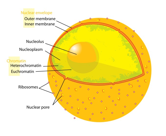
- The nuclear matrix or the nucleoplasm contains nucleolus and chromatin. The nucleoli are spherical structures present in the nucleoplasm. The content of nucleolus is continuous with the rest of the nucleoplasm as it is not a membrane bound structure. It is a site for active ribosomal RNA synthesis. Larger and more numerous nucleoli are present in cells actively carrying out protein synthesis.
- The interphase nucleus has a loose and indistinct network of nucleoprotein fibres called chromatin. But during different stages of cell division, cells show structured chromosomes in place of the nucleus.
- Chromatin contains DNA and some basic proteins called histones, some non-histone proteins and also RNA. A single human cell has approximately two metre long thread of DNA distributed among its forty six (twenty three pairs) chromosomes.
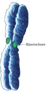
- Every chromosome essentially has a primary constriction or the centromere on the sides of which disc shaped structures called kinetochores are present.
- Based on the position of the centromere, the chromosomes can be classified into four types. The metacentric chromosome has middle centromere forming two equal arms of the chromosome. The sub-metacentric chromosome has centromere nearer to one end of the chromosome resulting into one shorter arm and one longer arm. In case of acrocentric chromosome the centromere is situated close to its end forming one extremely short and one very long arm, whereas the telocentric chromosome has a terminal centromere.
|
|
|
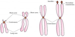
Sometimes a few chromosomes have non-staining secondary constrictions at a constant location. This gives the appearance of a small fragment called the satellite.
Karyotype
- It is the name given to the whole group of characteristics that allow the identification of a particular chromosomal set, i.e. the number of chromosomes, relative size, position of the centromere, length of the arms, secondary constrictions, and satellites.
- The karyotype is characterististic of an individual, species, genus, or larger grouping and may be represented by a diagram in which the pairs of homologues are ordered in a series of decreasing size.
- Some species may have special characteristics; for example, the mouse has acrocentric chromosomes, many amphibia have only metacentric chromosomes, and plants frequently have heterochromatic regions at the telomeres.
- Idiogram : The arrangement of chromosomes as per in order of their length, size, morphological features and diagrammatic representation of the haploid set of a species is called idiogram.
Special types of chromosomes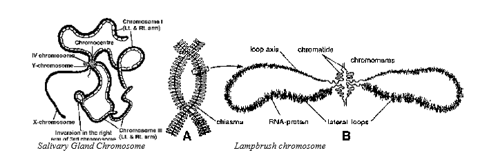
1. Polytene Chromosomes First seen by Balbiani (1881) in salivary glands of larva of Chironomus insect (midges — belongs to order Diptera).
- Term ‘Polytene’ by Koller (1888). Later on such chromosomes were observed in several other insects like Drosophila, mosquitoes.
- Polytene chromosomes are composed of many chromonema (512 to several thousands).
- Polyteny of giant chromosomes is achieved by replication of chromosomal DNA several times without nuclear division (endore duplication) and the resulting daughter chromatids do not separate but remain aligned side by side. These cells are unable to undergo mitosis and are destined to die during metamorphosis.
- In stained preparation, they appear striped with alternate dark and light bands. Dark bands have high amount of histone. Several puffy and swollen areas are seen on polytene chromosomes called as Balbiani rings or puffs. These are site of rapid m-RNA synthesis. Ecdysone hormone (moulting hormone) of insects stimulates formation of balbiani rings.
- Polytene chromosomes are concerned with metamorphosis in insects.
2. Lampbrush Chromosomes – Ruckert (1892). Seen in Oocytes of animals like shark, amphibians, reptiles and birds (most distinct in Diplotene stage). Also found in spermatocytes of several species, giant nucleus of Acetabularia and even in plants.
- Giant size of these chromosomes is due to increase in size of chromonema (not due to number). Chromonema grow in size to form lateral loops where synthesis of m-RNA occurs.
- Lampbrush Chromosomes are concerned with vitellogenesis (Yolk formation).
14. OTHER CELL ORGANELLES
Microbodies
Many membrane bound minute vesicles called microbodies that contain various enzymes, are present in both plant and animal cells.
- Single unit membrane bound, small spherical structures (9.3-1.5 µm in diameter).
- On the basis of functions, microbodies are of three types:
Spherosomes
- Hanstein (1880) first observed them and called as Perner (1953) named them spherosomes.
- Occur only in plant cells, and are major site of lipid storage and synthesis have lysosome-like activity, so also termed as plant lysosomes.
Peroxisomes
- In animal cells peroxisomes are concerned with peroxide metabolism. Urate oxidase, Amino acid oxidase and peroxidase catalyse the oxidation of amino acids producing
H. Catalase degrade the
into water and oxygen.
- In plants, peroxisomes occur in cells of green tissues and are concerned with photorespiration (glycolate pathway) and are also involved in -oxidation of fatty acids.
Glyoxysomes
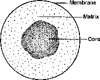
Structure of glyoxysome
- Occur only in plants especially in fatty seeds, guard cells of stomata and unripe fruits.
- Glyoxylic acid cycle takes place in Glyoxysome which was discovered by Kreb and Kornberg and is a variant of Kreb’s cycle.
Cytoskeleton
An elaborate network of filamentous proteinaceous structures present in the cytoplasm is collectively referred to as the cytoskeleton. The cytoskeleton in a cell are involved in many functions such as mechanical support, motility, maintenance of the shape of the cell.
These are fibrous or fine tubular structures which form the supportive structures of the cell. These are of two types — microtubules, microfilaments.
(a) Microtubules discovered by Robertis and Franchi, (1953) term coined by Slautterback (1963).
- Unbranched hollow non-contractile tubules of indefinite length, 25 nm in thickness with 15 nm core and formed of 13 helically arranged protofilaments of and -tubulin Microtubules grow from nucleating centres. Microtubules are basic structures of spindle apparatus, centrioles, basal bodies, cilia and flagella and are responsible for cell motility and maintenance of shape.
(b) Microfilaments are cylindrical solid, contractile rods or filaments of actin and myosin protein with a diameter of 6 to 10 nm. Microfilaments can form hexagonal bundles, take part in cytoplasmic streaming, membrane undulations, cleavage, contraction of muscles, movement of microvilli to absorb food and endocytosis.
Cilia and Flagella
- Cilia (sing.: cilium) and flagella (sing.: flagellum) are hair-like outgrowths of the cell membrane. Cilia are small structures which work like oars, causing the movement of either the cell or the surrounding fluid. Flagella are comparatively longer and responsible for cell movement.
- The prokaryotic bacteria also possess flagella but these are structurally different from that of the eukaryotic flagella.
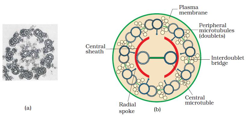
- The electron microscopic study of a cilium or the flagellum show that they are covered with plasma membrane. Their core called the axoneme, possesses a number of microtubules running parallel to the long axis. The axoneme usually has nine pairs of doublets of radially arranged peripheral microtubules, and a pair of centrally located microtubules. Such an arrangement of axonemal microtubules is referred to as the 9+2 array. The central tubules are connected by bridges and is also enclosed by a central sheath, which is connected to one of the tubules of each peripheral doublets by a radial spoke. Thus, there are nine radial spokes. The peripheral doublets are also interconnected by linkers. Both the cilium and flagellum emerge from centriole-like structure called the basal bodies.
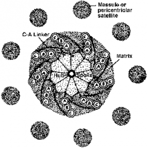
Centrosome and Centrioles
- Centrosome is an organelle usually containing two cylindrical structures called centrioles.
- They are surrounded by amorphous pericentriolar materials. Both the centrioles in a centrosome lie perpendicular to each other in which each has an organisation like the cartwheel.
- They are made up of nine evenly spaced peripheral fibrils of tubulin. Each of the peripheral fibril is a triplet.The adjacent triplets are also linked. The central part of the centriole is also proteinaceous and called the hub, which is connected with tubules of the peripheral triplets by radial spokes made of protein.
- The centrioles form the basal body of cilia or flagella, and spindle fibres that give rise to spindle apparatus during cell division in animal cells.