


1. TISSUE
A group of similar cells along with intercellular substances that usually have a common embryonic origin and function to carry out specialized activities. Body tissue can be classified into four principle types, according to their function and structure.
| Epithelial | Connective | Muscle | Nervous |
|
|
|
|
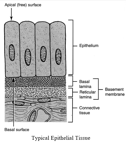
2. EPITHELIAL TISSUE AND ITS CLASSIFICATION
- Apical free surface which faces either body fluid or the outside environment.
- The cells are compactly packed with little intercellular matrix.
- Presence of basement membrane, except transitional epithelium.
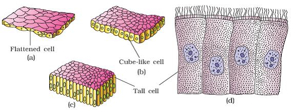
Based on cell layers and structural modification of the cells, epithelial tissue can be classified as –
| EPITHELIAL TISSUE | ||
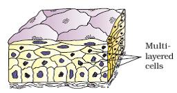
| Simple | Compound | Pseudo Stratified |
| Composed of single layer of cells which serves for absorption or filtration. | Composed of more than one layer of cells. Limited role in secretion and absorption, its main function is to provide protection against chemical and mechanical stress. | Cells of different sizes are arranged as single layer. |
Classification based on cell types, its arrangement and distribution.
Epithelial Tissues
| Type | Function | Location |
| Simple squamous epithelium | Filtration, diffusion, osmosis | Air sacs of lungs, walls of capillaries, lining of blood and lymph vessels. |
| Simple cuboidal epithelium. | Secretion, absorption | Surface of ovaries, lining of kidney tubules, and lining of ducts of certain glands |
| Simple columnar epithelium | Protection, secretion, absorption | lining of uterus and organs of the digestive tract |
| Pseudostratified columnar epithelium | Protection, secretion, movement of mucus and cells | Lining of respiratory passages and reproductive tract |
| Stratified squamous epithelium | Protection | Outer layer of skin, lining of oral cavity, throat, vagina, and anal canal |
| Stratified cuboidal epithelium | Protection, secretion | Male urethra, parts of the pharynx |
| Transitional epithelium | Distensibility, protection | Inner lining of urinary bladder and passageways of urinary tract |
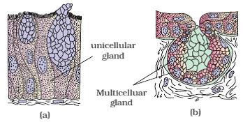
Glandular Epithelium
Composed of cells that are specialized to produce and secrete substances into ducts or into body fluids. Such cells are usually found in columnar or cuboidal epithelium.
| Glands – Based on number of cells | |
| Unicellular | Multicellular |
| e.g., Goblet cells of Alimentary canal. | e.g., Salivary glands, gastric gland, mammary gland. |
| Glands – Based on secretion | |
| Exocrine Gland | Endocrine Gland |
| Secrete their products into ducts that open into some internal or external surface. e.g. – Gland which secrete mucus, saliva, earwax, oil, milk, digestive enzymes and other cell products. |
Secrete their products into tissue fluid or blood. e.g. – Hormone secreting glands. |
Functional classification of exocrine gland.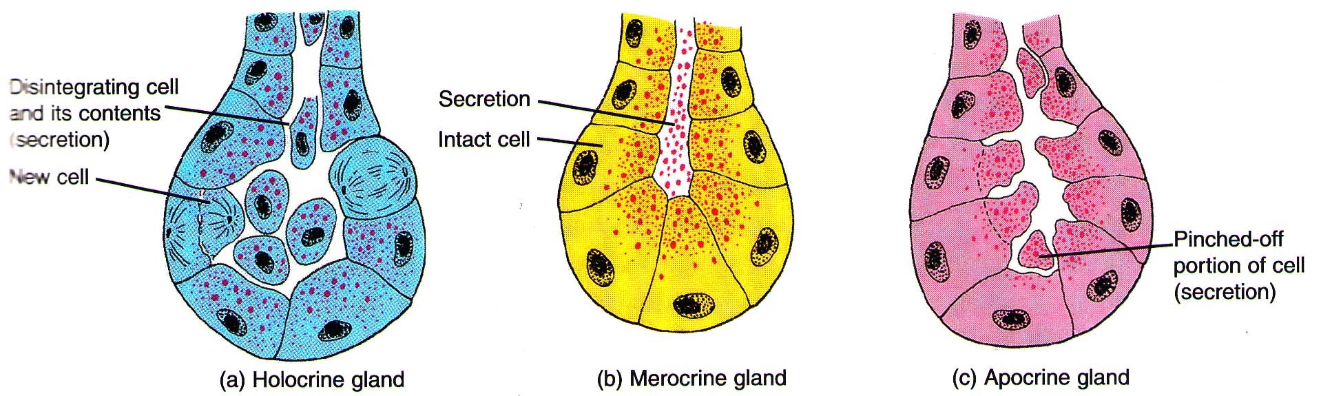
Holocrine gland: An entire cell when filled with secretory products, dies and disintegrates and is therefore discharged as a part of secretion (sebaceous gland).
Merocrine or epocrine gland: Secretion is discharged by the cells by simple diffusion so that there is no loss of the cells (most common exocrine gland). e.g., goblet cell, sweat gland, gastric gland
Apocrine gland: Secretory products accumulate in the apical parts of the cell, later this part breaks off from remaining part of a cell and is discharged as secretion. e.g., mammary gland
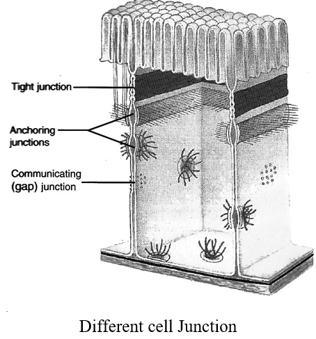
Cell Junction
Most epithelial cells, some muscle cells and some nerve cells are tightly joined to form a close functional unit. The points of contact between adjacent plasma membranes are called cell junctions.
| Tight Junction | Adhering or Anchoring Junction | Gap Junction |
| It forms fluid tight seals between cells which prevents substances from leaking across a tissue. | It performs cementing to keep neighbouring cells together and are common in tissues subjected to friction and stretching. | It provides cytoplasmic connection and allow electrical or chemical signals to pass from cell to cell. |
| Present between the cells of stomach, intestine and urinary bladder. | Outer layer of skin, muscle tissue of the heart. | Present in nervous tissue, muscle of the heart and gastrointestinal tract. |
3. CONNECTIVE TISSUE
This tissue provides the structural framework and support to different tissues, helps in body defense, tissue repair and fat storage etc. About 30% of the body mass is formed of connective tissue. It is most abundant and most widely distributed tissue in the body.
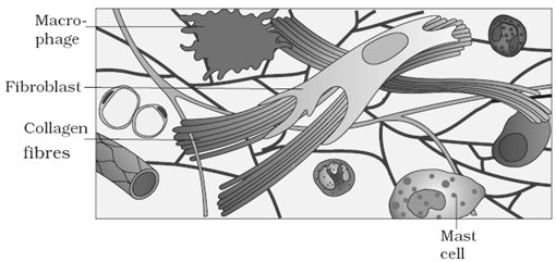
Components of Connective Tissue
Consists of various types of cells along with ground substance or matrix. It mainly consists of water and sulfated Mucopolysaccharides.
Components of connective tissue
| Component | Characteristic | Function |
| Fibroblasts | Widely distributed, large, star-shaped cells | Secrete proteins that becomes fibers |
| Macrophages | Motile cells that are sometimes attached to fibers | Engulf foreign particles from tissues by phagocytosis |
| Mast cells | Large cells, usually located near blood vessels | Releases substances that may help prevent blood clotting and promote inflammation |
| Collagenous fibers (white fibers) | Thick, threadlike fibers of collagen with great tensile strength | Hold structures together |
| Elastic fibers (yellow fibers) | Bundles of microfibrils composed of elastin that is very elastic | Provide elastic quality to parts that stretch |
| Reticular fibers | Thin fibers of collagen | Form supportive network within tissues |
Classification of Connective Tissue (CT)
| Connective Tissue | ||||||
| CT Proper | Supporting CT | Fluid CT | ||||
| Loose | Dense | Bone | Cartilage | Blood | lymph | |
| More matrix, less fibre e.g.–Areolar, adipose |
Regular | Irregular | ||||
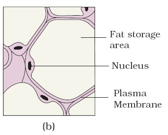
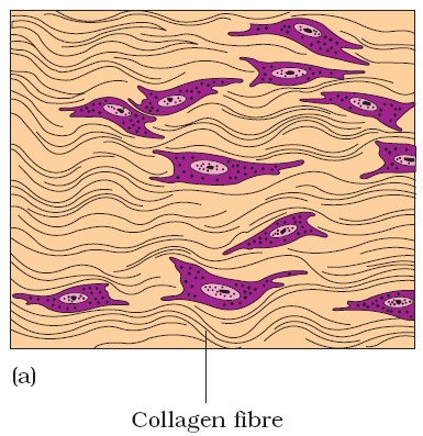
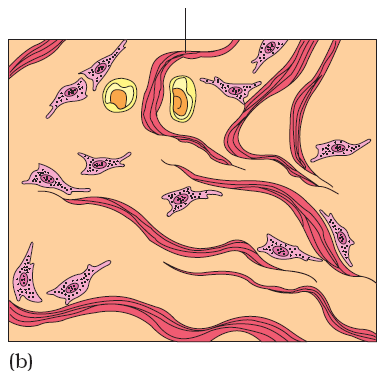
| Loose Connective Tissue Proper | |
| Areolar tissue | Adipose tissue |
|
|
| Dense Connective Tissue | ||
| Dense regular | Dense irregular | |
| Bundles of collagen fibres have a regular arrangement, in which fibroblasts appear in rows between the fibres. | It contains irregularly arranged collagen fibers and fibroblast cells.
|
|
| Tendon | Ligament | |
Formed of white fibrous connective tissue
|
Formed of yellow fibrous tissue, with collagen fibres
|
|
 |
 |
|
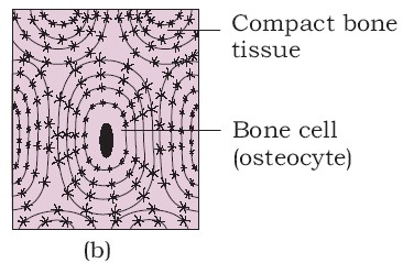
| SUPPORTING CT | |||
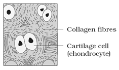
| Bones (Vascular) | Cartilage (Avascular) | ||
|
|
||
| Hyaline | Elastic | Fibrous | |
|
|
|
|
 |
 |
||
Fluid CT
Blood:
A type of liquid CT that helps in transportation, regulation (pH, body temperature) and protection (antibodies). It is composed of two parts approximately 55% is blood plasma and 45% is formed elements.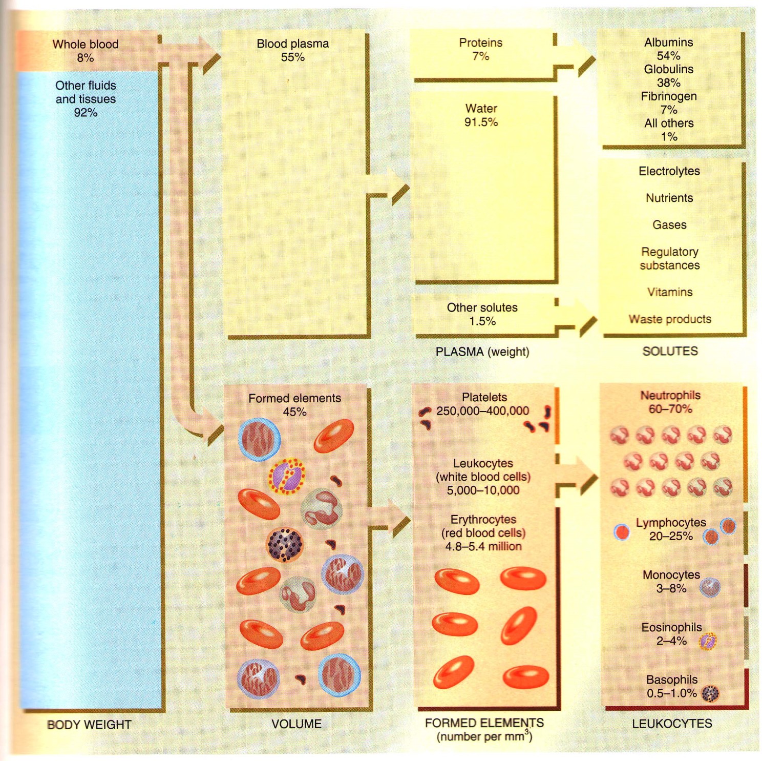
- Study of circulatory system is called as angiology. Father of blood circulation is William Harvey.
- Haematology is study of blood.
- Blood is the softest tissue in body and is derived from mesoderm.
- Blood is slightly alkaline (pH-7.4).
- Blood constitutes 8% of total body weight, blood volume in an average sized male is 5-6 litres and average female is 4-5 litres.
- pH of blood is maintained by balancing the ratio of sodium bicarbonate and carbonic acid. Buffer of blood is sodium bicarbonate.
- Blood is 2.5 times more viscous than water.
- Consists of watery fluid – plasma (55 to 60%), and formed elements – R.B.Cs, W.B.Cs, thrombocytes (40 to 45%).
- Plasma: Water (91-92%) solids (8-9%) out of which organic solids are 7% (albumin 4.4%, globulin 2.3%, fibrinogen 0.3%) and inorganic solids are 1%.
- Globulins are formed by plasma cells in lymphoid organs, other plasma proteins are formed in the liver.
- Globulins acts as antibodies. (immuno-globulins).
♦ R.B.C. (Erythrocytes)
- Amphibian R.B.C’s are largest amongst vertebrates, those of Amphiuma and Proteus are largest amongst amphibians.
- Mammals have smallest RBC’s amongst vertebrates. Musk deer have smallest amongst RBCs the mammals and elephant have largest RBCs among mammals.
- Whereas RBC’s of other vertebrates are oval and nucleated, those of mammals are roughly biconcave and without nucleus. This increases the surface area of RBCs and enables them to contain more haemoglobin.
- B.C count in Human – 5.4 million/mm3 of blood (slightly less in women, more in children).
- In embryonic condition, RBC’s are formed in mesoderm, yolk sac and bone marrow (haemopoietic organ).
- In adult mammal, red bone marrow in long bone is the main site for formation of RBCs.
- RBCs count increases during exercise and stress, and decreases during rest, sleep, menstruation, and pregnancy.
- Hill people have more RBCs. RBCs count sharply falls in anemia and rises in polycythemia.
♦ Life Span of RBC
- Life span of RBC in Rabbit – 80 days
- In man – 120 days
- In Frog – 100 days
- Radioactive chromium method. It is used for estimation of life span of RBC
ESR
- Erythrocyte sedimentation rate.
- It is measured by wintrobe’s tube. It is rate of settling down of RBC.
Haemocytometer
- It is an instrument used for counting both W.B.Cs. and RBCs.
Structure of RBCs of man
- Biconcave, non-nucleated bounded by Donnas membrane (plasma membrane of RBC)
- Donnan’s membrane is made of Lipoprotein which is 0.2 μ in thickness (mainly lipoid in nature)
- Lipid – 53% Protein – 40% Carbohydrate – 7%
W.B.C. Leukocytes
- Much larger than RBCs but less in number (about 8000/mm3 of blood).
- Ratio RBC : W.B.C. = 700 : 1
Two main types:
(1) Granulocytes (68%) : eosinophils, basophils, neutrophils
(2) Agranulocytes (32%) : lymphocytes, monocytes
(1) Granulocytes:
- Eosinophils (acidophils) – 2.8%, nucleus is An abnormal increase rise in its count is called eosinophilla which generally occurs in parasitic worm infestations. Stain dye is eosin. Number also increases in allergy condition and worm infection, e.g.– Ascaris. These are also known as Acidophils. They play an important role in hypersensitivity. These secrete histamine.
- Basophils – least numerous (0.5 to 2%), nucleus is nearly S shaped, functions like mast cells of connective tissue. Both basophil and eosinophil are involved in allergic reaction. Life span = 12-15 days. Nucleus is less distinct. They represent MAST Cells of connective tissue. Number increases in Chicken pox. These Secrete – (i) Histamine – Vaso dilater (ii) Heparin – Anti coagulant (iii) Serotonin – Vaso-constrictor.
- Neutrophils: most numerous (65%) and most active, nucleus 2 to 5 lobed. They engulf microbes and dead cells. Also known as Heterophils. Neutrophils are also known as micro policeman of blood (10 μ size). Life span = 2-4 days. Nucleus is multilobulated. Phagocytic in nature. Produce hydrolytic enzyme. Number increases in bacterial infection.
(2) Agranulocytes:
- Lymphocytes : 26%, nucleus large, produce antibodies. Small in size (7 μ ). Nucleus is central and rounded. Life span 3 days. Number increases in viral fever and whooping cough. Granules are absent in cytoplasm.
- Monocytes : 6%, nucleus-horse shoe shaped, phagocytic, changes into macrophages after entering tissue spaces. Largest WBC (22 μ ). It is macro policeman of blood. Life span = 28 days or one month. Number increases in tuberculosis (T.B.). They represent macrophages of connective tissue.
- Rise in W.B.C. Count is called
- Fall in W.B.C. Count is called leucopenia.
- Abnormal rise in W.B.C. Count is called
- TLC – Total Leucocyte Count = Normally 8000-9000 mm3
- DLC – Differential Leucocyte count
- Eosinophils – 2.8% Basophils – 0.2% Neutrophils – 65%
- Lymphocyte – 26% Monocyte – 6%
Blood Platelets
- Thrombocyte like cells.
- Found in mammals only.
- Platelets are derived from mega karyocytes in bone marrow.
- Life span = 10 days.
- Shape is irregular, oval of spherical.
- Platelets are non-nucleated.
- Count 2-5×10⁵ cells/mm3 or blood.
- Platelets are the fragments which form as bud and pinch off from certain large cell (mega karyocytes) in bone marrow.
- Blood platelets play an important role in blood clotting.
Spindle Cells or thrombocytes
- “Spindle cells” occur in all vertebrates other than mammals. Its function is like mammalian blood platelets.
- Nucleus is present, oval or spherical in shape.
- Help in blood clotting.
- Clot inside a vessel is called thrombus and its formation is known as
- Thalassemia is a inherited blood disorder resulting in anemia. The only treatment of thalassemia is Bone marrow transplant, has been performed for the first time in India at Christion Medical College Hospital in Vellore.
Life Span of Blood corpuscles
- R.B.C. (120 days in mammal, 100 days in frog), Granulocytes (1 to 8 hrs.) Agranulocytes (few days to a month), platelets – 8 to 10 days.
- ‘Spleen’ is often referred to as ‘graveyard’ of RBC (site of RBC disposal)
- W.B.C. mostly get destroyed in connective tissue.
Lymph:
White vascular connective tissue composed of plasma and leucocytes. It acts as a middle man between the blood and the tissue cells.
4. MUSCULAR TISSUE
Muscular Tissue
| Skeletal | Cardiac | Smooth | |
| Description | Long, cylindrical, striated fibers with many peripheral nuclei; voluntary control. | Branched cylindrical, striated fibers with one or two centrally located nuclei; contains intercalated discs; mainly involuntary control. | Spindle-shaped, nonstriated fibers with one centrally located nucleus; usually involuntary control. |
| Location | Usually attached to bones. | Heart wall | Walls of hollow internal structures such as blood vessels, airways to the lungs, stomach, intestine, gallbladder, and urinary bladder |
| Function | Motion, posture, heat production. | Pumps blood to all parts of the body die to their rhythmic contraction and relaxation. | Motion, constriction of blood vessels and airways; propulsion of food through gastrointestinal tract; contraction of urinary bladder and gallbladder. |
Skeletal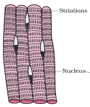 |
Cardiac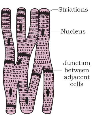 |
Smooth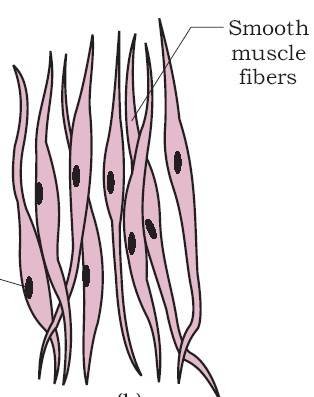 |
5. NEURAL TISSUE
It consists of only two principal kind of cells.
| Neural Tissue | ||
| Neurons or nerve cells | Neuroglia | |
| These can respond to stimuli and convert them into nerve impulses. | It makes up for more than one half the volume of neural tissue in our body. It protects and support the nerve cell. | |
| Location of Neuroglia in | ||
| CNS | PNS | |
|
|
|
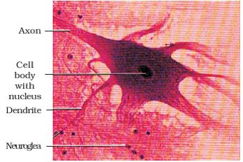
Neural tissue (Neuron with neuroglea)
ANIMAL MORPHOLOGY
6. EARTHWORM
Earthworm- (phylum-Annelida, Class-Oligochaeta) (Pheretima and Lumbricus)
- Reddish brown terrestrial invertebrates present in the upper layer of moist soil.
- Long, cylindrical body with 100-120 segments.
| BODY PART | |
| Dorsal surface | Ventral surface |
| Marked by a dark median mid dorsal line (dorsal blood vessel). | Distinguished by the presence of genital openings (pores). |
| Body Segments (Metameres) | ||
| 1st Peristomium | Clitellar (14,15,16) | Last |
| Bear prostomium, a lobe which serves as a covering for the mouth. | Dark band of glandular tissue, secretes cocoon. | Pygidium bears anus. |
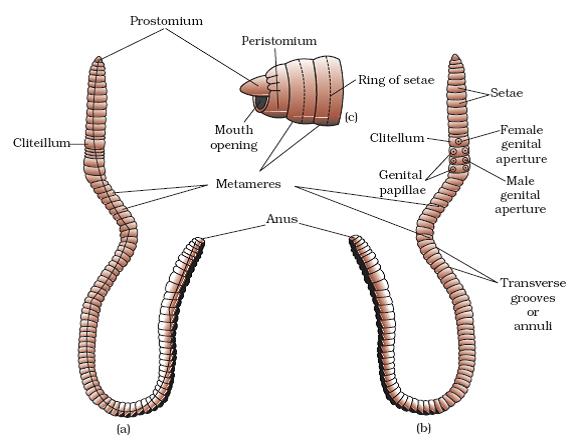
1 – 13 – Pre-Clitellar segments
14, 15, 16 – Clitellar segments
17 to last – Post clitellar segments
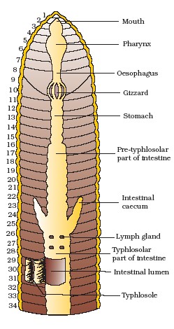
Body Segments and related parts
(1–3) → Buccal cavity
(4) → Pharynx
(5–7) → Oesophagus
(8–9) → Muscular gizzard → helps in grinding the soil particles and decaying leaves etc.
(9–14) Stomach → calciferous glands present in the stomach, neutralize the humic acid present in humus.
15 to last → Intestine
(15-26) → Pre typhlosolar
(27-95) → Typhlosolar
(95-Last) → Post typhlosolar
7, 9 → 2 pairs of lateral heart
12, 13 → 2 pairs of lateral oesophageal heart.
4, 5, 6 → Blood glands (Produce blood cells and haemoglobin which is dissolved in blood plasma.) Blood cells are also phagocytic in nature.
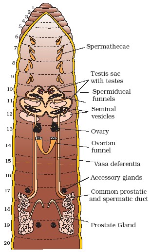
10, 11→ Two pairs of testes.
17, 19 → Two pairs of accessory glands.
6, 7, 8, 9 → Four pairs of spermathecae.
12, 13 → one pair of ovaries attached at the inter-segmental septum.
18th → A pair of male genital pores on the ventro-lateral side.
19th → A single median female genital pore.
BRIEF PHYSIOLOGY
Digestive system: Food (Humus ) is digested by saliva (with proteolytic enzyme) secreted by salivary gland cells, enzymes of intestinal caecae and epithelium of stomach and intestine. Digested food is absorbed in intestine, while undigested food and soil are egested through anus as worm castings.
Respiratory System: It lacks specialized breathing devices. Respiratory exchange occurs through moist body surface into their blood stream.
Circulatory System
- It exhibits a closed type of blood vascular system, consisting of blood vessels, capillaries, and heart.
- Dorsal blood vessel: Largest blood vessel provided with a pair of valves. It acts as collecting vessel (posterior to 13th segment as well distributing vessel (anterior to 13 segments).
- Ventral vessel: Principally a distributing blood vessel.
- Lateral-oesophageal Vessel: Collecting blood vessel.
- Supra-oesophageal Vessel: Collecting blood vessel.
- Lateral hearts: Connects dorsal to ventral blood vessel.
- Lateral oesophageal heart: It connects both dorsal and supra-oesophageal vessels with ventral vessel.
- Commissural vessel: behind 13th segment to last, it conveys blood from sub-neural to dorsal vessel.
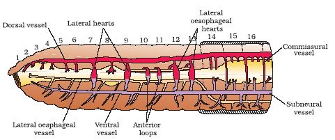
Closed circulatory system
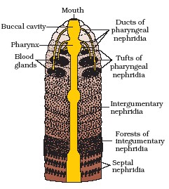
Excretory System: Takes place with the help of nephridia.
| Nephridia | |||
| Exonephric | Enteronephric | ||
| Integumentary | Pharyngeal | Septal | |
| Attached to lining of the body wall of segment 3 to the last. Open on the body surface through nephridiopores. | Three paired tufts in the 4th , 5th and 6th segments. | Present on both the sides of intersegmental septa of segment 15th to the last. It opens into intestine. | |
- A nephridium starts out as a funnel (nephrostome) that collects excess fluid from coelomic chamber. Nephridium delivers the wastes through a pore to the surface in the body wall or into the digestive tube.
- Nephridia regulate the volume and composition of the body fluids.
- Nephridia also help in osmoregulation.
Movement and Locomotion:
Locomotion takes place with the help of contraction and relaxation of body muscles and is aided by setae and coelomic fluid (acts as hydraulic skeleton).
Nervous System:
Represented by ganglia, arranged segment wise on the ventral paired nerve cord. The nerve cord in the anterior region (3rd and 4th segments) bifurcates, laterally encircling the pharynx and joins the cerebral ganglia dorsally to form a nerve ring. The cerebral ganglia along with other nerves in the ring integrate sensory input as well as command muscular responses of the body.
Sensory System:
Eyes absent but possess light and touch sensitive organs to distinguish the light intensities. To feel the vibrations in the ground specialized chemoreceptors (taste receptors) are located on the anterior part of the worm.
Reproductive system:
They are hermaphrodite (bisexual).
- A mutual exchange of sperm occurs between two worms during mating. Mature sperm, egg cells and nutritive fluid are deposited in cocoons produced by the gland cells of clitellum.
- External fertilization takes place in cocoons.
- Development is internal, takes place in cocoon.
- After 3 weeks each cocoon produces two to twenty baby worms with an average of four.
7. COCKROACH – Cockroach (Periplaneta americana)
Phylum – Arthropoda
Class – Insecta
- Brown or black bodied animals.
- Maybe bright yellow, red and green coloured.
- Nocturnal omnivores that live in damp places throughout the world.
- Segmented body with head, thorax, and abdomen.
- Complete body covered by hard chitinous exoskeleton.
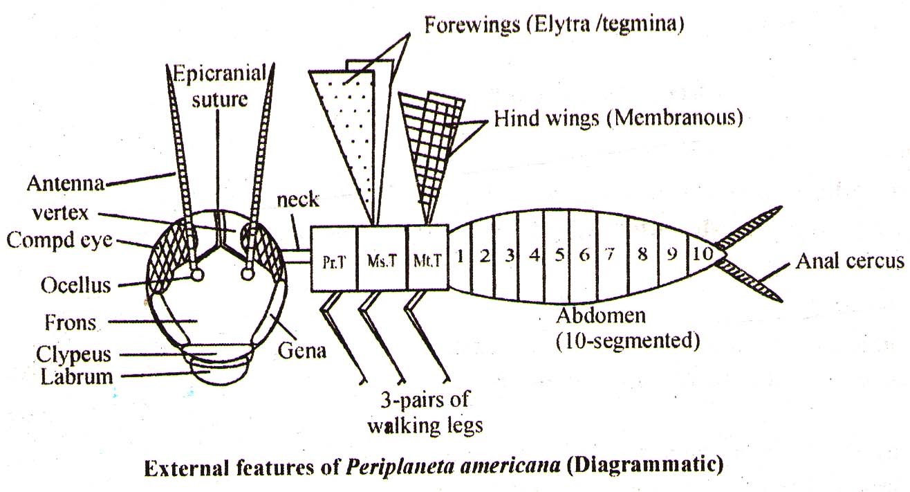
Head:
- Head consists of 6 fused segments and is downward directed (Hypognathous).
- Head bears one pair of segmented antennae (with sensory receptors that help in monitoring the environment).
- Presence of one pair of compound eyes and one pair of simple eyes.
- Presence of biting and chewing type mouth parts, which consist of upper lip (labrum), lower lip (labium) mandible, maxillae and hypopharynx (act as tongue).
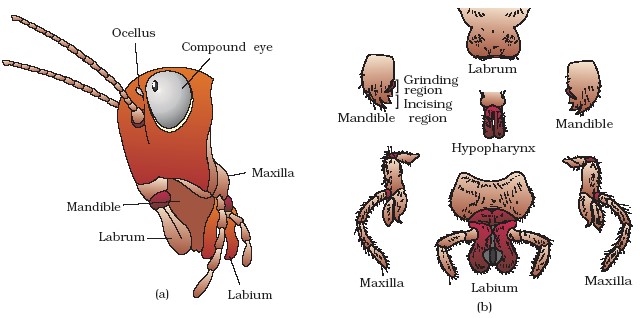
Head region of cockroach : (a) parts of head region (b) mouth parts
Thorax:
- It consists of three parts; prothorax mesothorax and metathorax.
- The exoskeleton of each segment is called sclerite (dorsal tergum and ventral sternum) are joined to each other, by thin and flexible articular membrane (arthrodial membrane).
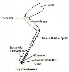
- Each thoracic segment has one pair of legs, this due to six legs they are called hexapoda.
- Meso and metathorax also bears wings. The metathoracic (forewings) called tegmina are opaque, dark, and leathery and cover the hind wings when at rest. Metathoracic hindwing) is membranous and is used in flight.
- Abdomen:
Ten segmented in both male and female.
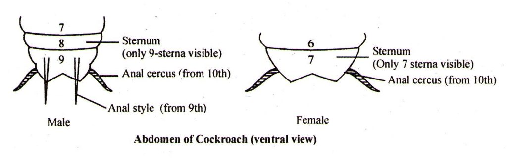
| Male | Female |
|
|
Brief anatomy and physiology:
Digestive system:
- Alimentary canal consists of 3-regions-foregut (stomodaeum), midgut (mesenteron) and hindgut (proctodaeum).
- Foregut and hindgut are internally lined with cuticle.
- Crop: digestion and storage of food.
- Gizzard: Masticating organ (made up of 6-cuticular plates and circular muscles).
- Digestion takes place in the crop and midgut, with the help of saliva (from one pair of salivary glands) and enzymes from hepatic caeca and midgut cells.
- Absorption takes place in midgut and hindgut (water and mineral).
- Hepatic caeca (8) at the junction of foregut and midgut.
- Malpighian tubule at the junction of midgut and hindgut.
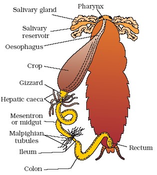
Alimentary canal of cockroach
Respiratory system (Tracheal system)
- It consists of a network of trachea that open through 10 pairs of small openings called spiracles, present on the lateral side of the body.
- Each tracheal tube is internally lined with cuticle called intima which prevents collapsing of the tracheal tubes.
- Branches of trachea carry oxygen from the air to all the parts of the body. Exchange of gases take place at the tracheoles level by diffusion.
Circulatory System:
- Open type.
- A tubular 13 – chambered, segmented heart , located in pericardial cavity and suspended with the help 12 pairs of alary muscles.
- Blood vessels are poorly developed and open into space (haemocoel). Visceral organs located in the haemocoel are bathed in haemolymph.
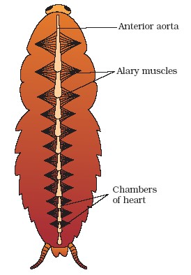
- Haemolymph is composed of colourless plasma and haemocytes.
- Except last chamber, all other chambers have one pair of ostia (12 pairs) guarded with valves.
- Due to peristaltic wave of heart chamber haemolymph moves from posterior to anterior direction.
- Haemolymph has little to do with oxygen transport in most insects, it helps in the transportation of excretory and nutritive materials only.
Excretory System:
- Primary excretory organ → (50 – 60) Malpighian tubule.
- Accessory excretory organ → Fat body, Nephrocytes and Uricose glands.
- Malpighian tubule is lined by glandular and ciliated cells. It absorbs nitrogenous waste products and convert them into uric acid, which is excreted out through the hindgut (uricotelic).
Nervous System:
- It consists of a series of fused, segmentally arranged ganglia joined by paired longitudinal connective on the ventral side.
- There are three thoracic and six abdominal ganglia.
- Small part of nervous system is present in the head, while the rest is situated along the ventral part of its body.
- Supra-oesophageal ganglia represents the brain.
Sensory Organs:
It includes antennae, eyes, maxillary palps, labial palps, anal cerci etc.
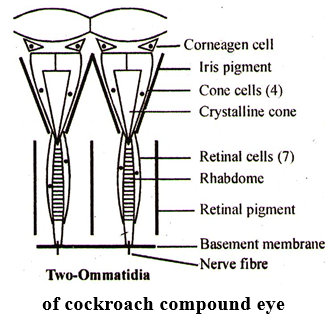
- It includes highly developed one pair of compound eye. The functional unit of compound eye is hexagonal ommatidium (2000 in each compound eye). Such eye forms many images in pieces with the help of several ommatidia. Such image is called apposition mosaic image with more sensitivity but less resolution.
- Apposition (non-overlapping) image for diurnal insect (except cockroach)
- Super position image (nocturnal insect).
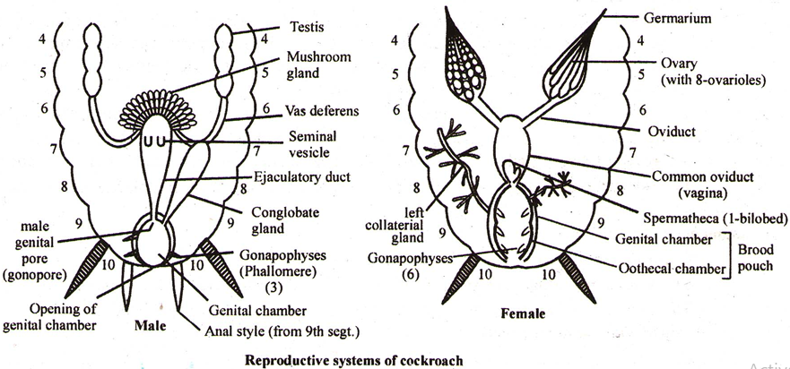
Reproductive System:
- They are dioecious with distinct sexual dimorphism.
- Male reproductive system includes a pair of testes (4–6 abdominal segment).
- A pair of vas deferens.
- Single ejaculatory duct and gonopore.
- Three gonapophysis (Phallomere).
- A pair of seminal vesicles.
- A mushroom shaped gland (6-7 abdominal segment).
- Female reproductive system includes:
- A pair of ovaries (2–6 segment).
- A pair of oviducts.
- Single vagina, gonopore, and spermatheca.
- 3 pairs of gonapophysis.
- one pair of collateral glands.
Mechanism of reproduction:
- Each ovary is formed of a group of eight ovarian tubules or ovarioles, containing a chain of developing ova. Each ovariole forms one mature ovum at one time. Sixteen ova are conducted into genital chamber.
- Sperms are stored in the seminal vesicle and are glued together in the form of bundles called spermatophores, which are discharged during copulation (by left phallomere) in the female spermatheca.
- Sperms are released from spermathecae hence internal fertilization takes place.
- Secretion of collateral glands forms ootheca around fertilized eggs.
- Oothecas are dropped at some safe place (more humid and near a food source).
- On an average female produce 9–10 oothecae, each containing 14 – 16 eggs.
Paurometabolous development:
- The nymphs look very much like adults. The nymph grows by moulting about 13 times to reach the adult form. The next to last nymphal stage has wing pads but only adult cockroaches have wings.
8. FROG – Phylum- Chordata
Class-Amphib
- Rana tigrina is commonly called as Indian Bull- frog
- It is amphibious and can be found in ponds/ shallow water bodies and in terrestrial conditions.
- It is cold blooded or Poikilothermic and shows summer –sleep (Aestivation) and winter –sleep (Hibernation) in extreme temperatures.
- Its body colouration shows camouflage. Its colour changing ability is called Metachrosis. This protective colouration is called mimicry.
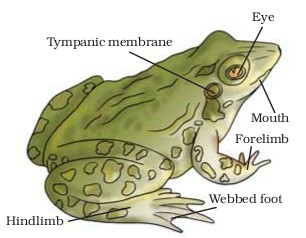
External morphology-
- Its skin is slimy moist without exoskeleton or scales.
- The body of the frog is divisible into head and trunk. The trunk has one-pair of shorter fore limbs and one pair of longer hind limbs. Each fore limb has 4-fingers (digit) whereas each foot has 5-toes(digit). (The thumb, pollex, or the first finger in fore limb is absent in frog)
- Eyes are dorso-laterally placed on head. Frog has 3-eye lids, i.e. upper eye lid , lower eye lid and third eye lid (nictitating membrane), the later covering the eye-ball during swimming.
- External ear is absent in frog. The tympanum / ear drum is present near the eyes.
- Frogs exhibit sexual dimorphism. Male frogs can be distinguished by the presence of sound producing vocal sacs and a copulatory pad on the first digit of the fore limbs which are absent in female frogs.
Internal morphology (anatomy) and physiology-
Digestive system-
- Due to carnivorous nature, the alimentary canal of frog is short. The posterior most part of gut is cloaca. Urine also passes through cloacal opening.
- Due to absence of Uvula, the buccal cavity and pharynx are not differentiated. The bucco-pharynx contains numerous maxillary teeth. The teeth in frog are Homodont, Acrodont and Polyphyodont. The teeth are however absent in lower-jaw. Besides upper-jaw, the teeth are also present in vomer bone of olfactory capsule in the roof of bucco-pharynx. The teeth in frog are not used for cutting the food material but are used for preventing the escape of the prey. The tongue in frog is bifid at the tip and is free behind. It is used for capturing prey (insects).
- Presence of liver which secretes bile, and pancreas secretes digestive enzymes.
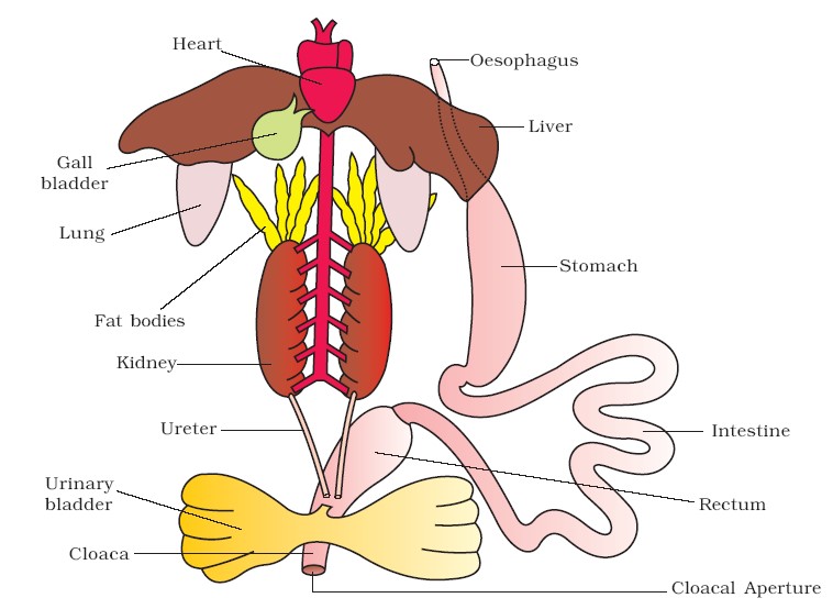
Diagrammatic representation of internal organs of frog showing complete digestive system
Respiratory system-
- Frog respires through skin (Cutaneous respiration) bucco –pharyngeal lining and through lungs (Pulmonary respiration). During aestivation and hibernation, the only mode of respiration is cutaneous.
- The lungs are a pair of elongated, pink coloured sac-like structures present in the upper part of the trunk region (thorax). Air enters through the nostrils into the buccal cavity and then to lungs. The lungs are hollow, non-lobular and positive pressure type.
Blood vascular system
- In frog BCS is closed type and has double circulation through not as efficient as in birds and mammals.
- The heart is 3-chambered (2-atria and 1-ventricle). The dorsal side of the heart has a triangular structure, called Sinus venosous which receives deoxygenated blood from 2-precavals and 1-postcaval. It pours the blood into right atrium. On the ventral side of the heart there is a prominent structure, called conus arteriosus which arises from the ventricle.
- The blood from the heart is carried to all parts of the body by the arteries (arterial system). The veins collect blood from different parts of body to the heart and form the venous system.
- The SA node is present in the wall of sinus venosus and not in the right atrium as in mammals.
- There are 2-types of portal systems in the body of frog, i.e. Hepatic portal system (between gut and liver) and Renal portal system (between posterior abdominal parts and kidney).
- Lymphatic system consists of lymphatic hearts. Lymphatic vessel, and lymphatic capillaries.
Nervous system
- Frog has all 3-types of nervous system, i.e. CNS (brain and spinal cord), PNS (Cranial nerves and spinal nerves) and ANS (SNS and PSNS).
- Brain is enclosed in a bony structure called brain box (cranium). The brain is divided into forebrain, midbrain, and hindbrain. Forebrain includes olfactory lobes, paired cerebral hemispheres and unpaired diencephalon.
The midbrain is characterised by a pair of optic lobes. Hindbrain consists of cerebellum and medulla oblongata. The medulla oblongata passes out through the foramen magnum and continues into spinal cord, which is enclosed in the vertebral column. - The cranial and spinal nerves in frog are 10-pairs each. However, in Rana tigrina the number of spinal nerves is 9-pairs.
Sensory organs
- Frog has different types of sense organs, namely organs of touch (sensory papillae), taste (taste buds), smell (nasal epithelium), vision (eyes) and hearing (tympanum with internal ears).
- The eyeball in frog is cartilaginous. The colour vision is lacking due to the absence of cones in retina. In place of lachrymal and meibomian glands, there are Harderian glands in frog.
- External ear is absent in frogs and only tympanum can be seen externally. The ear is an organ of hearing as well as balancing (equilibrium).
Excretory system
- The excretory system consists of a pair of kidneys, ureters, cloaca, and urinary bladder.
- Each kidney is composed of several structural and functional units called uriniferous tubules or nephrons.
- The kidneys in frog are mesonephros. Such kidney is not differentiated into cortex and medulla.
- Each kidney has 4-5 pairs of renal artery and veins.
- The nephrons or uriniferous tubules are not differentiated into PCT, DCT and Henle’s loop.
- Two ureters emerge from the kidneys in the male frogs. The ureters act as urinogenital duct which opens into the cloaca. In females the ureters and oviduct open separately in the cloaca.
- The major excretory product in frog is urea (Ureotelic).
Reproductive system
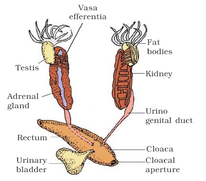
(i) Male reproductive system
- The testes are attached to kidney through fold of peritoneum, called mesorchium.
- The sperms from testes pass through vas efferentia (10-12 in number) and are released into Bidder’s canal, a collecting duct of the kidney. Finally, it communicates with the urinogenital duct that comes out of the kidneys and opens into the cloaca. The cloaca is a small, median chamber that is used to pass faecal matter, urine, and sperms to the exterior.
(ii) Female reproductive system
- The ovaries are also present close to the kidney, but unlike testes, are not functionally connected to it. The oviducts open separately into cloaca.
 |
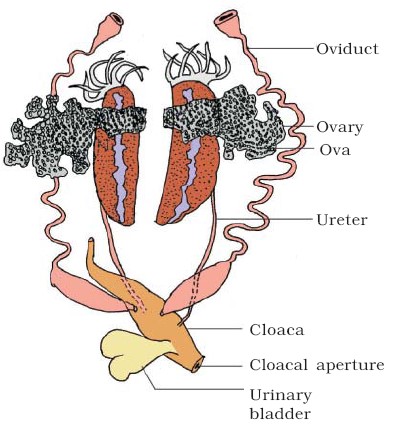 |
| Male reproductive system | Female reproductive system |
Development
- The Frog is spontaneous or seasonal ovulator, i.e. the Ovulation occurs according to the season. The eggs (2500-3000) are laid in spawn.
- The copulation (without copulatory organ in male) is false and is called Amplexus. The Fertilization is external (in water) and cleavage is unequal holoblastic.
- The development is indirect through tadpole larvae.
Metamorphosis
- It occurs in the presence of thyroxine hormone which changes larvae into adult frog. In case of deficiency of thyroxine or iodine, the tadpole increases in size and does not undergo metamorphosis.