


1.1 CELL
Have you ever seen a cell?
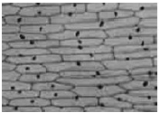
Take an onion bulb. Peel off the skin from its inner side using forceps. Then, place individual layers in a watch glass containing water. Carefully transfer each layer on a clean glass slide using a paint brush. Add one-two drops of iodine solution on the material to stain the cell for visualization. Place a cover slip on the material in such a way that no bubbles are formed. Observe the slide under the microscope.
What do you observe?
You will observe a number of rectangular chambers under the microscope (as shown in the given figure). Each chamber is a cell here. You will find a similar pattern and structure in each slide.
Cell and its Naming
All living organisms are made up of cells. The Cell is the structural and functional unit of life. It is the smallest living entity found in living organisms.The term cell was introduced by Robert Hooke in 1665,while examining a slice of cork through his microscope.Cork is obtained from the bark of a tree. Under a compound microscope, he observed many small compartments resembling a honeycomb. He termed these
as cells.
Historical Account
1. The simple microscope was built by Zacharias Janssen and was modified by Galileo. The first compound microscope was made by Robert Hooke (1665). He examined thin slices of cork under his microscope and observed the honey comb like structures composed of box like compartments which were termed as the cellulae (cells). His work was published in his book “Micrographia”. Cells were observed prior to Hooke by Malpighi (1661), who called them saccules and utricles.
2. Leeuwenhoek (1674) observed few living cells capable of moving, such as bacteria, protozoa,spermatozoa and red blood corpuscles under his own designed microscope.
3. Lamarck remarked that “no living being can have life if its constituent parts are not formed by cells”.
4. In 1831, Robert Brown discovered the presence of nucleus in the cells of orchid root. Fontana discovered nucleolus in the skin cell of Eel. The term nucleolus was given by Bowman.
Properties of cell
The Cell is the smallest living unit of life.
It is so small that it is not visible to the naked eye.
The shape of the cell varies in different organisms and within an organism.
Size of cells also differs.
All living cells exhibit certain basic properties like respiration, growth, metabolism etc.
Cells originate from a pre-existing cell. A mother cell divides to produce daughter cells. Hence, cells exhibit cell division.Cell is the basic unit of life. All cells vary in their shape, size, and activity they perform. In fact, the shape and size of the cell is related to the specific functions they perform.
The table given below lists the shape and size of certain cells.
|
Cell |
Shape |
Size |
|
Bacteria Human egg Muscle cell Human epidermal cell Red blood cells White blood cells Lymphocytes
|
Round shape, rod shape, spiral shape etc. Round Rod-shaped with pointed ends Columnar Bi-concave Irregular Round shaped |
0.1 to 0.5 μm 100 μm in diameter 1 – 40 mm long 7 μm in diameter 10 12 μm 5 6 μm in diameter |
Evolving of multicellular organisms
Multicellular organisms have several advantages over unicellular organisms.
As division of labour exists in multicellular organisms, a variety of tasks can be performed efficiently. This
gives the organism a wide range of adaptability to survive.
In multicellular organisms, dead cells play an important role. For example, dead epidermal cells in the skin of
animals protect the underlying cells.
Division of labour
Division of labour suggests the specialized functions of organs. All organs, tissues, or cells cannot carry out all the
functions. They are evolved to carry out a specific set of functions. Each organ system coordinates with the other to carry out functions required for life. Therefore, by dividing the work or function, they minimize the load of carrying out all functions and therefore, they work or function efficiently.
Let us learn more about unicellular and multicellular cells. Organisms are often groupedas unicellular or multicellular organisms. Unicellular organisms represent a single cell, while multicellular organisms are made up of numerous cells.
|
Unicellular organism |
Multicellular organism |
|
A single cell represents an organism. The entire organism is made up of a single cell. Characteristics 1. A single cell represents an organism. 2· The cell or organism is independent and does not need help from other such cells for its growth and development. 3· There is no division of labour present in a unicellular organism. 4· They avail nutrients from the environment. 5· The cell can change its shape according to the environment. 6· They usually reproduce by binary fission. |
More than one or many cells combine and coordinate to form a multicellular organism. Characteristics 1· An organism is composed of numerous cells. 2· Cells in multicellular organisms depend upon other cells present in the organism for their growth and development. 3· There is division of labour in multicellular organisms. Specific cells carry out their specific functions. 4· They avail nutrients by the intake of food. 5· Cells in multicellular organisms exhibit definite shape. For example, neuron has a definite and distinct shape, which they do not change. 6· They reproduce through sexual and asexual modes. |
|
Few examples of unicellular organisms are bacteria, virus, yeast, Amoeba, Paramecium, Chlamydomona etc. |
Few examples of multicellular organisms are humans, plants, insects, snakes, birds, lizards etc. |
Some Interesting Facts
The smallest cell in the universe is the Mycoplasma, a type of bacteria. Its diameter is 0.1 μm.
The smallest cell in the human body, in terms of volume, is the sperm cell.
The table given below lists the characteristics of some unicellular and multicellular organisms with a few
examples.
Cells can also be classified on the basis of their cellular complexity.
Based on their sub-cellular organization and cellular complexity, cells can be classified as prokaryotes and eukaryotes.
Animals, plants, fungi, protozoans, and algae, all are composed of eukaryotic type of cells, while bacteria are prokaryotes in nature.
Prokaryotes are unicellular organisms, while eukaryotes are usually multicellular organisms. Yeast is exceptionally a unicellular eukaryote. The table given below lists the characteristic features of both prokaryotes and eukaryotes
|
Characteristic |
Prokaryote |
Eukaryote |
|
Size of the cell |
Small in size |
Vary in size, generally larger than prokaryotes |
|
Nucleus |
Nucleus with nuclear membrane is absent |
A well-defined nucleus with nuclear membrane is present |
|
Membrane -enclosed organelles |
Organelles like mitochondria and plastids, which have a membrane around them are absent |
Membrane-enclosed organelles like mitochondria and plastids are present |
|
Cell wall |
Cell wall is usually present in prokaryotes and is composed of peptidoglycan |
Cell wall is usually present in plant cells and is composed of cellulose |
|
Genetic material |
Present as nucleoid |
Present inside the nucleus |
1.2 STRUCTURE OF THE EUKARYOTIC CELL
These are cells having organised nucleus and two envelope system e.g., protista, fungi, plantae and animalia. The subcellular components of the cell are: cell wall, plasma membrane, cytoplasm, mitochondria, chloroplast, Golgi body, nucleus, cytoskeleton, lysosome and other cell organelles. The cytoplasm is the main arena of cellular activities in both the plant and animal cells.
1.3 CELL WALL
It is the outer rigid and dead protective covering of plant cells, majority of prokaryotes and fungi. The cell wall
was first observed by Robert Hooke in 1665 in a thin slice of cork. Certain cells, such as gametes and zoospores, lack cell wall. The cell wall varies in thickness from 0.1 m to 10 m in different cells. The cell wall is secreted by the cytoplasm.
Chemical Composition of Cell Wall
Algae have cell wall, made of cellulose, galactans,mannans and minerals like CaCO3, while in other plants it consists of cellulose, hemicellulose, pectins and proteins.
Water: 30-60%, Hemicellulose: 5-15%, Pectin
substances: 2-8%, Lipid: 0.5-3%, Proteins: 1-2%,
Microfibrils: 20-40% and deposition: 0-25%
Layers of Cell Wall
(a) Middle lamella
(b) Primary cell wall
(c) Secondary cell wall
(a) Middle lamella: It is intercellular matrix (common layer) between the primary walls of adjacent cells.
It is thin amorphous cementing layer composed of pectates of calcium and magnesium. It is the first
structure formed from cell plate between newly formed daughter cells at the time of cytokinesis.
Ripe fruits get softened due to partial solubilisation of pectic compounds which give jelly like
consistency.
(b) Primary cell wall: It is first formed wall and is capable of growth. It develops in the growing cells
such as immature meristematic and parenchymatous cells on the inner side of middle
lamella. It is formed of pectates, cellulose, hemicellulose and some polysaccharides. It is thin,
amorphous, elastic, permeable, 0.1 to 0.3 m thick layer which grows by incorporation of more wall
material within the existing one. Such a growth is called intussusception.
(c) Secondary cell wall: When the plant cell enlarges with the development of large vacuole, the
secondary cell wall appears inner to primary wall.It is thick (3-10 m) rigid, composed of compact
macrofibrils of cellulose in which the matrix is formed of cellulose, hemicellulose and other
polysaccharides impregnated with lignin, suberin,cutin, silica, calcium and magnesium salts, waxes,
resins, tannins and gums. Lignin forms hard wood and suberin forms cork. Growth pattern is called
accretion, e.g., Sclerenchyma.
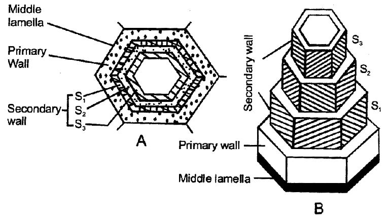
Cell wall layers: (A) A cell in T. S. showing parts of cells wall. (B) Typical wood fibre cut to show parts and
layers of the secondary wall.In some tissue a tertiary cell wall is formed on the inner surface of the secondary cell wall. This layer is very thin and is found in the xylem tracheids of gymnosperms. It is composed of xylan and
mannan.
Functions of Cell Wall
The cell wall performs the following important
functions:
(i) It provides a definite shape, protection and
mechanical support to the cell.
(ii) It functions as apoplast.
(iii) It forms a skeletal framework of plants and provide
mechanical support.
(iv) Its depositions like cutin, and suberin reduce
transpiration.
(v) It is involved in the movements of metabolites in
and out of the cell through plasmodesmata.
(vi) It counteracts the turgor pressure.
1.4 CELL MEMBRANE
Cell membrane is thin, elastic, semifluid, dynamic, living protective membrane which occurs inside and
outside the cytoplasm of the cells. The term ‘cell membrane’ was given by Nageli and Cramer. Inside the cytoplasm of eukaryotic cells, it covers various cell organelles like-mitochondria, vacuole, plastids, microbodies, etc.
These membranes are biologically similar, dynamic and interconvertible.
They help in compartmentalisation of the cells because of their property of selective permeability.
The plasma membrane (PM) or Plasmalemma form the outermost boundary of cytoplasm which separates
it from the extracellular environment and controls the entrance and exit of molecules and ions and thus helps
to maintain the difference in ion concentration of the cytoplasm and the surroundings.
The term ‘plasmalemma’ was coined by Plowe (1931).
Structure of cell Membrane
The detailed structure of the membrane was studied only after the advent of the electron microscope in the 1950s.
Chemical studies on cell membrane, especially in human RBCs, enabled the scientists to deduce the possible
structure of plasma membrane.Under electron microscope plasma membrane appears as a tripartite structure consisting of two parallel dark lines separated by a central clear area. The two parallel dark lines were found to be layers of protein molecules whereas, a central clear area is a layer of biphospholipid layer. Both lipid layers remain linked with each other by the inner ends of lipid molecules which are non-polar and hydrophobic in nature.
This ensures that the non polar tail of saturated hydrocarbons is protected from the aqueous environment. The lipid molecules remain linked with the molecules of protein layers by their hydrophilic ends.
The protein layers provide elasticity and mechanical
Later, biochemical investigation clearly revealed that the cell membranes also possess protein and
Carbohydrate.
The ratio of protein and lipid varies considerably in different cell types. In human beings, the membrane of the erythrocyte has approximately 52 per cent protein and 40 per cent lipids.
|
FUNCTIONS OF PLASMA MEMBRANE |
|
· It protects intracellular organelles from the outside environment. · It selectively allows the molecules to move in and out of the cell. Gases such as carbon dioxide and oxygen pass through diffusion, while other small molecules such as sugar pass through a process called passive transport. Ions such as sodium or potassium are transported through a process called active transport (with the utilization of ATP). Movement of water molecules through the membrane takes place by osmosis. · It can also engulf or expel substances in and out of the cellthrough endocytosis or exocytosis respectively. Endocytosis is the process through which cells take in extra-cellular substances. Endocytosis represents both phagocytosis (ingestion of whole food),and pinocytosis (ingestion of water). · It establishes communication between cells. |
Diffusion
In the digestive tract, food is broken down into simpler products such as glucose. Nutrients are then absorbed
by the intestinal cells through a process known as diffusion.
The inhaled air contains oxygen. In the lungs, oxygen diffuses through the blood capillaries and reaches the
red blood cells. There, it binds with haemoglobin to form oxy-haemoglobin. Oxy-haemoglobin is then
circulated throughout the body. In tissues, where oxygen concentration is lesser than blood or the red blood cells,oxygen diffuses out of it and enters into tissues and then into cells.
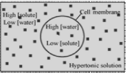
Osmosis
Osmosis is the movement of water molecules across a semi-permeable membrane from a higher to a lower
concentration.Plasma membrane acts as a semi-permeable membrane.Water moves in and out of cells through osmosis to maintain the amount of water in cells and in the outside environment.
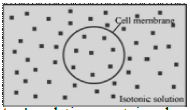
Solutions can be of three types: isotonic, hypotonic, and hypertonic. Let us explore the differences between these solutions.
An isotonic solution contains similar solute (salt or sugar) and water concentration, both inside the cell and in the medium (where the cell is suspended). While working with biological samples, sa lt solut ion of 0.9% is used as an isotonic solution.
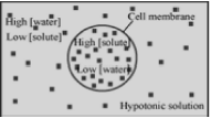
A hypotonic solution contains lesser salt (i.e., higher water concentration) in the medium than the cell. The cell contains
higher solute and lower water concentration.When cells are kept in such a solution, water enters inside the cells. It results in
swelling and bursting of cells.
A hypertonic solution conta ins more salt concentration ( i.e., lower water concentration) than cells. When cells are placed in such solutions , water moves out of the cells. The cell shrinks , resulting in the breaking down of the plasma membrane.
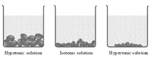
Let us perform an experiment to understand osmosis using raisins or apricots.
Take raisins in three breakers. Pour pure water in the first beaker, isotonic solution in the second, and a high concentrated salt solution in the third beaker.
What do you observe?
After five minutes, you will observe that the raisins swell up in the first beaker. Pure water does not contain salt. Thus, to maintain equilibrium, water enters into the raisins. This swells up the raisins.
In the second beaker, you will see no change in the raisins. An isotonic solution contains similar salt concentration as that of raisins. This produces osmotic balance. Hence, no change can be observed.
In the third beaker, you will observe that the raisins shrink. This beaker contains a hypertonic solution. To maintain equilibrium, water moves out of the raisins.
This shrinks the raisins. Excessive shrinkage results in the breakdown of the membrane. This phenomenon is called plasmolysis.
Do you Know | Artificial kidney dialysis!
An artificial dialysis must be performed when the kidneys stop functioning. Artificial kidney dialysis uses the cellulose membrane, which acts as a semi-permeable membrane. Such a system filters blood through osmosis and diffusion (as the kidneys do). This keeps the body activity normal.
|
Diffusion |
Osmosis |
|
Solute molecules move across a concentration gradient. |
Water molecules move across a concentration gradient . |
|
It does not require a semi-permeable membrane. |
It requires a semipermeable membrane. |
Plasma membrane of cells acts as semi-permeable membrane. It allows the movement of water and gaseous molecules freely (diffusion). However, it does not allow the movement of other larger molecules such as sugar,amino acids, etc. All such molecules are transported across the membrane by facilitated diffusion (with the help of other carrier proteins) and active transport (with the expenditure of energy).
Significance of diffusion in living organisms
|
Biological importance of diffusion |
|
· Gaseous exchange in the lungs over the surface of alveoli: Oxygen from air diffuses into blood while carbon dioxide gas diffuses out of blood. · Gaseous exchange in tissues: Oxygen from blood diffuses out and enters the tissues. Carbon dioxide gas produced as waste is removed by diffusion. |
Significance of osmosis in living organisms
|
Biological importance of osmosis |
|
· Plant roots absorb soil water through osmosis. Water concentration is high in soil as compared to root cells. Therefore, water moves from soil to cells. · Water is re-absorbed in the proximal and distal convoluted tubules of nephron (kidney) through osmosis. · Water is absorbed in the digestive tract through osmosis. Upon drinking, water concentration becomes high in the digestive tract as compared to the cellular water concentration. |
Do you know why brine is used to preserve food?
Brine is a high salt concentration solution. Perishable food items such as fish, meat, etc. are preserved for a longer period of time in such solutions. Brine is hypertonic for bacteria. Therefore, water comes out of bacterial cells, causing dehydration. This kills the bacteria. Thus, bacterial contamination is prevented.
Importance of plasma membrane
1. The cell membrane maintains the individuality and form of the cell and its organelles.
2. A cell remains dynamic as long as the plasma membrane is able to determine which material should enter or leave the cell. Cell membrane helps in regulating the flow of materials and energy into and out of the cell through the processes of diffusion, osmosis and diffusion.
3. It helps to form sub-cellular organelles within the cytoplasm.
4. It controls the cellular interactions essential for the tissue formation and defence against the entry of microbes.
5. Its glycoproteins and glycolipids (glycocalyx) play a distinct role in blood grouping, immune responses, etc.
6. Peripheral proteins in cell membrane have enzymatic activities and also make the membrane selectively permeable.
7. Cell membrane plays a big role in cellular movements by the formation of pseudopodia, cilia and flagella.
8. Plasma membrane helps in the exit of secretions and wastes by exocytosis.
9. Cell membrane has receptors for certain hormones.
10. Membrane infolds are used for bulk intake of materials by endocytosis.
1.5 CYTOPLASM
The cytoplasm is the inner content of the cell membrane, which is separated from the nucleus. It includes cytosol, organelles, and inclusions. Cytosol is a soft and sticky, semi-transparent fluid in which various cell organelles are suspended. Inclusions are stored nutrients.
The table given below lists various functions of the cytoplasm.
Functions of Cytoplasm
It is the region where many cellular activities take place.
It mostly consists of water and it balances the water content in the cell.
It contains cytoskeleton, which maintains the shape and movement of cells.
Cytoplasmic streaming or circulation of the cytoplasm helps in the proper distribution of cellular organelles during cell division, growth, etc.
Do you know what cybrid is?
Cybrids are cytoplasmic hybrids. Plasma membrane of cells (of different origins) is broken down to obtain cytoplasm. These naked cells are then fused to obtain hybrid cells called cybrids. Cybrids are often known as heterokaryon as they contain the nucleus from different origins. Cybrids are important for research purposes.
Some Interesting Facts
· Cells can be grown outside the body through a process known as cell culture.
· Cell culture media is always isotonic in nature. An isotonic solution has salt concentration of 0.9% of NaCl, which equals to the intracellular salt concentration.· Viruses do not have plasma
membrane and cytoplasm.
ENDOMEMBRANE SYSTEM
Cell have extensive sets of intracellular membranes, which together compose the endomembrane system. It was first discovered by Camillo Golgi. The functions of this system are coordinated. The endomembrane system includes the endosplasmic reticulum (ER), Golgi complex, lysosomes and vacuoles. Membrane components, including proteins and lipids, are exchanged among these organelles and the plasma membrane via vesicular transport. Since the functions of mitochondria, chloroplast and peroxisomes are not coordinated with the above components, these are not considered as part of the endomembrane system.
1.6 ENDOPLASMIC RETICULUM
The endoplasmic reticulum was first noticed independently by Porter and Thompson. It was named ER by Porter. The cytoplasm in eukaryotic cells is traversed by a complex network of inter-connecting membrane bound structures. They divide interacellular space in two compartments, i.e., luminal (inside ER) and extra luminal (cytoplasm). These can extend from nuclear envelope to plasma membrane.
Ultrastructure
The endoplasmic reticulum is composed of the following three kinds of structures, viz., cisternae, vesicles and tubules.
Cisternae
The cisternae are long, flattened, parallel, sac-like, interconnected structures. These are found in cells which are actively involved in protein synthesis. The cisternae usually occur in those cells which have synthetic roles, e.g., cells of pancreas and brain.
Tubules
The tubules are branched or unbranched structures forming the reticular system alongwith the cisternae and vesicles. They are free of ribosomes and are common in cells involved in lipid and sterol synthesis.
Vesicles
The vesicles are oval, membrane bound vacuolar structures. They are free of ribosomes. They are abundant in the pancreatic cells and these are only ER structures found in spermatocytes.
Types of ER
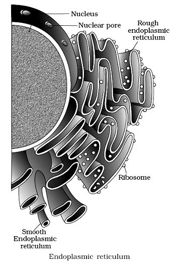
There are two types of endoplasmic reticulum.
1. Agranular or Smooth Endoplasmic Reticulum (SER).
This type of endoplasmic reticulum possesses smooth walls because the ribosomes are not attached with its membranes. The smooth endoplasmic reticulum is generally found in adipose cells, interstitial cells, glycogen storing cells of the liver, spermatocytes and leucocytes. The muscle cells are also rich in smooth types of endoplasmic reticulum and are known as sarcoplasmic reticulum.
2. Granular or Rough Endoplasmic Reticulum (RER).
The granular or rough type of endoplasmic reticulum possesses rough walls because the ribosomes remain attached with its membranes, with the help of proteins called ribophorin I and II. The rough type of endoplasmic reticulum is found abundantly in those cells which are active in protein synthesis such as pancreatic cells and liver cells.
Common Functions of Smooth and Rough ER
Provides support to the colloidal protoplasm (Endoskeleton).
Transport and exchange of materials.
Localization of organelles: It keeps the cell organelles properly distributed in relation to one another.
Formation of desmotubules which extends through the plasmodesmata.
Functions of Smooth ER
Smooth ER synthesizes fats and lipids.
It also takes part in the metabolism of Carbohydrates.
It actively participates in drug detoxification.
It maintains the calcium ion concentration in the cytosol.
Functions of Rough ER
Most of the lysosomal proteins are produced in the rough ER.
It transports proteins to various destinations like the plasma membrane.
This is the major site of glycosylation (addition of Carbohydrates in proteins).
1.7 GOLGI APPARATUS
Golgi apparatus are the membrane-bound, sac-like structures called cisternae. They are arranged parallel to each other in stacks. They were discovered by Camillo Golgi in 1898. Golgi body is usually composed of fiveeight cisternae in stacks. Vesicles leave the Golgi body from one end known as the cis face. The other end is known as the trans face. The table given below lists
some functions of the Golgi apparatus.
Structure
It is surrounded by a zone of cytoplasm which is devoid of organelles. It is called zone of exclusion. Golgi complex is in the form of a parallelly arranged interconnecting system of a cluster of smooth membranes having a central stack of flattened sacs or cisternae (size: 0.5 mto 1.0 m diameter), with many peripheral tubules, and vesicles lying above the nucleus, around or above the centriole in animal cells and scattered in cytoplasmic matrix in plant cells. Golgi complex has two distinct faces i.e., forming face (cis region) and a maturing face (trans region).
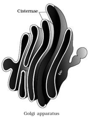
There are four parts of the Golgi complex:
(a) Cisternae: These appear in the form of flat, curved, smooth membraned structures with swollen ends. They have a definite polarity. Their convex pole or proximal pole or cis face is associated with nuclear envelope. The concave pole or distal pole or trans face, is the oldest, which gives rise to the secretory vesicles.
(b) Tubules: These are small, flat, interconnecting structures arising from the periphery of cisternae due to fenestrations.
(c) Vesicles: These are large rounded sacs present at the edges of cisternae in clusters. These are pinched off from the tubules.
(d) Golgian vacuoles: These are large, spherical vacuoles produced at maturing face. These are filled with some granular or amorphous substances. Some of them function as lysosomes. Functions of Golgi apparatus
The main function of Golgi apparatus is to process, package, transport and release of secretory proteins. Materials to be packaged in the form of vesicles from the ER fuse with cis face of the Golgi apparatus and moves towards the maturing face.
This explains, while the Golgi apparatus remains in close association with the ER. A number of proteins synthesised by ribosomes on the ER are modified in the cisternae of the Golgi apparatus before they are released from its trans face.
They cause glycosidatioin of lipids and glycosylation of proteins to form glycolipids and glycoproteins.
Acrosome of the sperm is modified Golgi apparatus.
Root cap cells are rich in Golgi bodies which secrete mucilage for the lubrication of root tip.
Golgi apparatus helps in the recycling of broken plasma membrane during endocytosis.
Most of the proteins synthesised at ER are modified
in Golgi bodies.
1.8 LYSOSOMES
Christian de Duve (1955) is credited with the discovery of lysosomes. These are single membrane bound small vesicular structures rich in hydrolyzing enzymes (acid hydrolases).
Ultrastructue
Lysosome is surrounded by a single unit membrane. it encloses a dense stroma and a large vacuole. The vacuole contains about 50 hydrolytic enzymes (acid hydrolases) which can digest most of the biological substances. Hydrolytic enzymes of lysosomes act at
acidic pH. The acidic condition is maintained by pumping protons into the interior of lysosomes. Some of the hydrolytic enzymes are acid phosphatases, acid ribonucleases, acid deoxyribonucleases, cathepsin, glycosidases, etc.
This organelle shows polymorphism: On the basis of morphology, their contents and functions, lysosomes are divided into following four forms:
1. Primary lysosomes: These are small, vesicle-like newly formed structures produced from the Golgi apparatus, at trans face. Primary lysosomes contain inactive enzymes.
2. Secondary lysosomes: These are also called heterophagosomes or digestive vacuoles.
Secondary lysosomes are formed when phagosomes fuse with already existing primary lysosomes. These contain the enzymes against the material to be digested.
3. Residual bodies: The secondary lysosomes accumulate undigested substances. They are secondary lysosomes formed from digestive and autophagic vacuoles which contain only undigested materials. Residual bodies pass outwardly, come
in contact with plasmalemma and throw their contents to the outside through ephagy or exocytosis. However, in certain cells the residual bodies do not discharge their contents to the
outside. Instead, they load the cells and bring about
ageing, e.g., liver cells, muscle cells.
4. Autophagic vacuoles: They are formed by union of many primary lysosomes around old or dead organelles, surround them with vacuolar membrane and digest them by autolysis or autodigestion.
Origin
Lysosomes arises from bud off vesicles of the Golgi complex at the trans face.
Functions
(i) Extra and intracellular digestion.
(ii) Acrosome part of sperms release enzymes to dissolve the egg membranes for the entry of the sperm into the ovum during fertilisation.
(iii) Certain diseases are caused by nondigestion of certain components due to absence of required enzymes in the lysosomes. Most of the diseases result from accumulation of glycolipids and mucopolysaccharides.
(iv) Accidental or pathological release of hydrolases from lysosomes causes breakage of chromosomes, their abnormal distribution during mitosis and mutations. Cancer is also caused by such a change.
1.9 MICRO-BODIES
These are single membrane bound organelles associated with oxidation reactions other than those of respiration.
These include:
(i) Sphaerosomes
(ii) Peroxisomes
(iii) Glyosysomes
Sphaerosomes
They are also called as half membrane bound organelle. These were discovered by Perner. These are actually unit membrane bound, spherical, refractile bodies which take part in storage and synthesis of fats. These are in abundance in endosperm cells of oil seeds. Sphaerosomes arise from smooth endoplasmic reticulum. These contain hydrolytic enzymes. These are believed to be plant lysosomes.
Peroxisomes
Peroxisomes are spherical, sac-like structures, bounded by a single membrane. Initially these were reported in animals only, but later also discovered in plants.
De Duve discovered them for the first time. These were called peroxisomes, because these contain ‘peroxide’- producing enzymes (oxidases) and ‘peroxide’-destroying enzymes (catalases).
The oxidative enzymes include –and –hydroxy acid oxidase, D-amino acid oxidase, and urate oxidase.
These catalyse the oxidation of substrates producing . Catalase degrades to water and oxygen.
is a by product of several biochemical reactions.
Since it is a powerful oxidising agent, it is extremely toxic to the cell.
All green cells of plants contain peroxisomes. These generally arise from ER. In plants, peroxisomes perform photorespiration. (Tolbert et. al. 1969). These contain the enzyme glycotate oxidase associated with photorespiraton.
Glyoxysomes
Glyoxysomes were discovered by Tolbert and Beevers. These originate from ER and are bounded by a single membrane. These contain enzymes for the glyoxylate cycle through which fats are converted into Carbohydrates (gluconeogenesis).
These are found in germinating seeds, especially in germinating fatty seeds (such as castor seeds, ground
nut seed, etc.), where insoluble lipid food reserves must be turned into soluble sugars for the growing tip.
Animals cannot execute this conversion because they do not possess glyoxylate enzymes.
1.10 MITOCHONDRIA
Mitochondria were discovered by Kolliker (1880) in striated muscles of insects and the present name was given by Benda (1897). Altman called mitochondria as bioplasts. Mitochondria are stained with Janus green B – a vital stain. They are absent in prokaryotes. Mitochondria are secondarily lost in the red blood corpuscles of mammals. Mitochondria are present in the living eukaryotic cells.
Size: Commonly mitochondria are 1.0-4.1 min length
and 0.2-1.0 m (average 0.5 m) in diameter.
Shape: The common shape is cylindrical or sausageshaped.
It is spherical in yeast. Other forms are filamentous, club-shaped, racket-shaped or vesicular.
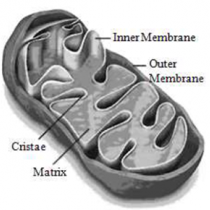
Mitochondrion is a membrane-enclosed organelle found in eukaryotic cells. A mitochondrion is composed of two lipid membranes, enclosing the matrix. The inner membrane gets folded to form numerous cristae. Cristae are the main site for ATP production. Mitochondrial matrix contains mitochondrial DNA and ribosomes.
Mitochondria are responsible for the production of most of the energy (or ATP) in cells. Therefore, mitochondria are also known as the power house of cells.
Functions of mitochondria
Mitochondria are the power houses of the cell.
These bring about the oxidation of Carbohydrates, proteins and -oxidation of fats.
These are site of aerobic respiration, where Krebs cycle occurs in matrix, while ETS and oxidative phosphorylation enzymes are located in inner membrane.
Mitochondria are the sites for the formation of certain intermediates which are important in the synthesis of chlorophyll, cytochromes, pyrimidines, steroids, alkaloids, etc.
These help in the synthesis and elongation of fatty acids.
Synthesis of many amino acids occurs in mitochondria.
Mitochondrial inheritance, independent of nuclear inheritance is found in some cases, petite character in yeast and cytoplasmic male sterility in maize are examples of mitochondrial inheritance.
1.11 PLASTIDS
E. Haeckel (1865) gave the term plastid. Plastids are largest cell organelles. These are involved in the formaton and storage of soluble and insoluble Carbohydrates.
Plastid inheritance, independent of nuclear inheritance
has been observed in Mirabilis jalapa (4 o’clock plant).Plastidome is the sum total of plastids (carrying genetic
information) in a cell.
Schimper discovered different types of plastids. These can be divided in three types on the basis of presence or absence of different pigments:
1. Chloroplast
2. Leucoplast
3. Chromoplast
Chloroplast
Schimper gave the term chloroplast.
Morphology: Shape, size and number of chloroplasts vary from species to species. A variety of shapes can be observed. specifically among green algae. In higher plants, chloroplast may be ovoid, spheroid or discshaped but mostly they are Iens-shaped.
Size: Generally the size ranges in length (5 – 10 m) and width (2 – 4 m).
Structure
Each chloroplast is bounded by two unit membranes separated by space called periplastidial space. Outer membrane contains integral proteins called porins. Inner membrane is selectively permeable, having carrier proteins for transport. Parts inner to membrane is divided into two portions:
(i) Grana, (ii) Stroma
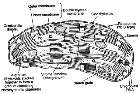
(i) Grana: Grana fraction consists of a membranous or lamellar system. This lamellar system is made up of thylakoids (term given by Menke). Light reaction of photosynthesis occurs in this fraction. Agranal chloroplast is found in bundle sheath cells of plants.
(a)Smaller thylakoids are placed one above the other in form of stacks of coins and form a granum.
The number of grana per chloroplast is 40-60 and the number of thylakoids per granum is 2-100. On the inner side of thylakoid membrane is present the Iumen. There are present minute granular structures called quantasomes embeded in thylakoid membrane which are morphological expressions of photosynthetic (light reaction) units.
Park and Biggins discovered quantasomes. Each quantasome has 230 chlorophyll molecules and about 50 caroteniids.
(b)Larger thylakoids are also present between two grana, these are long membranous structures which are named as stroma lamellae or fret channesls.
(ii) Stroma: Dark reaction of photosynthesis occurs in this region. The stroma is proteinaceous complex.
It has 2-6 copies of circular double stranded DNA.
Ribosomes are 70 S type. Enzymes of Calvin cycle (specially Rubisco), lipid metabolism and sugar interconversion are present here. Chloroplast also shows semi-autonomous nature.
Functions:
The chloroplasts perform various functions like;
1. Photosynthesis – light reaction (in thylakoids),Dark reaction (in stroma). The main function of chloroplast is photosynthesis, in which radiant energy of sun is converted into chemical form of energy, which is utilized by all living organisms to perform their life activities. Further, chloroplasts
help in maintaining balance of and in the atmosphere.
(2) Storage of starch.
Leucoplasts These are colourless plastids found in storage organs away from sunlight. Granum is absent e.g., underground stem or root. These are generally rod-shaped or circular. Leucoplasts are of three types
(a) Aleuroplasts or proteinoplasts: These store proteins, e.g., in maize.
(b) Elaioplasts: These store lipids or fats, e.g., in endosperm cells of castor seeds.
(c) Amyloplasts: These store starch, e.g., in potato tubers, wheat grains.
Chromoplasts
These are non photosynthetic plastids and are variously coloured (except green) providing orange, brown colour to flowers, in some young leaves, ripened fruits as well as in algae. These stores carotenoid pigments.
These different types of plastids are interchangeable.e.g., In tomato
Young ovary (colourless) – Leucoplasts
Young fruits (green) – Chloroplasts
Mature fruits (red) – Chromoplasts
In carrot root: Leucoplast – Chromoplast
In chilli: Chloroplast – Chromoplast
1.12 RIBOSOMES
The ribosomes are submicroscopic, non membrane bound granular particles of the ribonucleoprotein. The ribosomes were first noted in plant cells by Robinson and Brown with the electron microscope and Gorge Palade (1953) observed them in animal cells.
Occurrence and Distribution: The ribosomes occur in all cells. In prokaryotic cells, the ribosomes often occur freely in the cytoplasm. In eukaryotic cells, the cytosolic ribosomes either occur freely in the cytoplasm or remain attached to the outer surface of the endoplasmic reticulum (80 S) and organelle ribosomes inside mitochondria and chloroplast (70 S).
Structure
The ribosomes are composed of two subunits. One ribosomal subunit is large in size and has a dome-like shape, while the other ribosomal subunit is smaller in size and occurs above the larger subunit and forms a cap-like structure. These are negatively charged molecules containing rRNA and protein.
The two ribosomal subunits remain united with each other due to a specific concentration of the ions. When the concentration of ions reduces below a critical level both ribosomal subunits get separated. But, by raising the concentration of ions in the matrix, the two ribosomes become associated with each other and is known as the dimer. During protein synthesis, many ribosomes form a chain on a common messenger RNA and form the polyribosomes or polysomes, or ergasome.
Origin
In prokaryotes, both ribosomal RNA and proteins are synthesized in the cytoplasm. In eukaryotes, ribosomal RNA (hence ribosomal sub units) is synthesized in the nucleolus.
Functions
(i) The free ribosomes synthesize non-secretory proteins, while ER bound ribosomes synthesize secretory proteins.
(ii) The proper folding of proteins is assisted by specific proteins called as Chaperons.
1.13 NUCLEUS
The nucleus was discovered by Robert Brown (1831) in the root cells of an orchid. J. Hammerling (1953) conducted experiments on a single-celled green alga, Acetabularia and proved hereditary role of nucleus in morphogenesis.
It is double membrane bound dense protoplasmic body that controls cellular metabolism, encloses all the genetic information and is able to transmit the same to the next generation. Hence, it is also called as master organelle.Nucleus of undividing cell is often called interphase nucleus.
Nucleus is present in all the eukaryotic cells. Some cells contain nucleus when they are young but it degenerates when cells mature, e.g., sieve tubes in plants and RBC in mammals (anucleate cell). In prokaryotic cells ‘true nucleus’ is absent.
Generally, there is one nucleus in each cell, but this number may vary. Depending upon the number of nuclei, cells are called uni–, bi– or multi-nucleate. Multinucleate condition may arise either by fusion of may cells (syncytium) or due to repeated nuclear divisions with failure of septa formation (e.g., coenocyte). The shape and size of the nucleus varies with the type and the function of the cell. It may be rounded, oval or disc like.
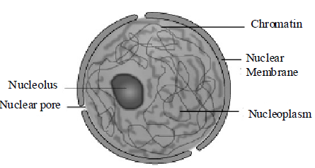
Structure
The nucleus can easily be distinguished into following
four parts.
(a) Nuclear membrane
(b) Nucleolus
(c) Nucleoplasm
(d) Chromatin
(a) Nuclear Membrane or Nucleolemma or karyotheca: It forms the envelope of the nucleus, present in all the eukaryotic nuclei, except during late prophase of the cell division. In prokaryotic cells, nuclear membrane is always absent.
Nuclear membrane consists of two unit membranes.
The space between the two membranes is called perinuclear space (10 – 50 nm). The outer unit membrane is continuous with endoplasmic reticulum, sometimes having attached cytoplasmic ribosomes (80 S). The inner membrane is smooth at its both surfaces. The nuclear membrane is interrupted by pores. These are called nuclear openings or pores. They maintain continuity between nucleo-cytoplasmic regions. Nuclear pores, are plugged by a cylinder of protein material called annulus, both collectively form pore complex. The passage of ions and small molecules through the pores is regulated by annulus.
Nuclear membrane allows a free exchange of ions.
Nuclear pores are the passages through which movement of RNA and protein molecules takes place in both directions between the nucleus and the cytoplasm.
Nuclear lamina is a network of acidic proteins and intermediate filaments, present just inside the inner nuclear membrane. It is also called fibrous lamina. This forms attachment site for telomeres during leptotene stage of meiotic prophase I.
(b) Nucleolus: This organelle was first discovered by Fontana and the term nucleolus was given by Bowman. It is spherical, dense, colloidal, acidophilic body that remains attached to a special type of chromosome having a specific position called Nucleolar Organising Region (NOR) or secondary constriction. Larger and more numerous nucleoli are present in cells actively carrying out protein synthesis.
(c) Nucleoplasm: The nucleus is filled with a transparent, semifluid, granular substance, known as nuclear sap or nucleoplasm or karyolymph.
Nucleoplasm is mainly made of nucleic acids, proteins, enzymes, lipids and minerals. The nucleic acids –DNA and RNA are found in association with histone proteins and acidic proteins or non-histone proteins. The important nuclear enzymes include
DNA polymerase, RNA polymerase, nucleoside phosphorylase etc. Minerals like phosphorus, sodium, and magnesium are also present.
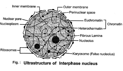
(d) Chromatin: Chromatin (Flemming) is the hereditary part of the nucleus. It occurs as a uncondensed network of filaments or fine threads. During prophase stage of cell division, it organises
itself in the form of chromosomes. It is differentiated into two regions.
Heterochromatin and Euchromatin: It was observed that when chromosomes are stained with basic dyes like acetocarmine or fuelgen stain, then two types of regions can be observed:
(i) Heterochromatic region: This region gets dark stain during interphase. This is genetically inactive and highly condensed region with tightly packed DNA.
(ii) Euchromatin region: This region gets light stain during interphase. This portion is genetically active and rich in loosely packed DNA.Transcription occurs here.
Chromosome
The chromosomes were discovered by Hofmeister and were named as chromosome by Waldeyer.
The shape of chromosomes depend upon the position of
centromere. Common shapes on the basis of position of centromere are follows:
(i) Metacentric: Here the centromere is situated in the median position of chromosome producing two equal arms. These chromosomes appear V-shaped.
(ii) Sub-metacentric: In such type of chromosomes, the centromere is slightly displaced from the centre, forming one long and another slightly shorter arm.
These chromosomes appear L shaped.
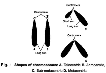
(iii) Acrocentric: When the centromere is located subterminally, forming a very short and a long arm.
These appear J shaped.
(iv) Telocentric: Here the centromere is situated at one
end of the chromosome (terminal). These appear ‘i’ shaped.
If centromere is absent, the chromosome is known as acentric, and if there are two centromeres it is called dicentric.
Structure of Chromosome
Structurally, a chromosome is composed of following parts:
1. Chromatid: Each metaphase chromosome consists of two symmetrical strands called chromatids.
2. Chromonema: Termed by Vejdovsky, during prophase each chromatid appears to be made of very thin and highly coiled filaments called chromonemata (as subunits of chromatids).
3. Chromomeres: These are bead-like structures formed due to condensation of chromatin material. These are believed to be location of genes.
4. Centromere: The two chromatids are attached to each other by a narrow area, also called primary constriction. This decides the ratio of arm length called centromeric index. Two arms may be equal (lsobrachial) or unequal (Heterobrachial).
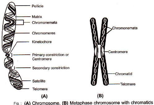
5. Kinetochore: This is a disc-shaped protein structure attached to the centromeric portion.
6. Telomere: This term is applied to the cytologically distinct tips of the chromosomes, these are specific for a chromosome and are rich in G and T bases.
7. Nucleolar Organising Region (NOR): These areas are certain secondary constrictions containing the genes which code for ribosomal RNA that induce the formation of nucleoli. The chromosomes with these regions are called nucleolar organising
chromosomes.
8. Satellites: This is a rounded body separated from the rest of the chromosome by a secondary constriction, a chromosome having satellite is called SAT-chromosome and these are considered marker chromosome (SAT – Sine Acid Thymonuclenico).
Depending upon sex and other body characters, the chromosomes in eukaryotes are of two type:
(i) Autosomes: These are more in number and have nothing to do with sex, but determine other body characters.
(ii) Sex chromosomes or Heterosomes or ldiosomes:
These chromosomes are concerned with sex. These are commonly of ‘X’ and ‘Y’ type.
Functions of chromosomes
(1) Chromosomes are responsible for carrying the genetic information from one generation to another, as genes are located on them.
(2) Any variation in chromosome (structural or numerical) will lead to change in the characters of an organism.
1.14 VACUOLES
These are non cytoplasmic areas present inside the cytoplasm which are separated from the latter by specific membranes. These are believed to be formed by expansion and pinching off from ER. In plant cells, the vacuoles can occupy upto 90% of the volume of the cell.
Depending upon the contents and their functions, vacuoles are of 4 types:
1. Sap Vacuoles: These are filled with fluid matrix (cell sap) and are bounded by a unit membrane called tonoplast. In plants, tonoplast facilitates the transport of a number of ions and other materials against concentration gradients into the vacuole,
hence their concentration is significantly higher in the vacuole than in the cytoplasm. These have water soluble pigments called anthocyanin and anthoxanthin, responsible for colour of petals. It also contains mineral salts, sugars, amino acids, esters, waste material, etc., This maintains OP of a cell. Alkaloids and tannins are also stored here.
2. Contractile Vacuoles: These are found in fresh water protozoans and algal cells. These take part in osmoregulation and excretion.
3. Food Vacuoles: These are formed by the fusion of a phagosome and primary lysosome in some protozoans and phagocytes of higher animals.These contain digestive enzymes and help in the digestion of nutrients.
4. Gas Vacuoles (Air or Pseudovacuoles): These are
found in some prokaryotes only and contain metabolic gases. These protect the cells against harmful radiations and regulate buoyancy of the cell.
1.15 CELL INCLUSIONS
These are metabolically inactive, non-living substances present in the cell, also called ergastic substances. these can be organic or inorganic in nature and may get solubilized or remain insoluble.
These include reserve materials, excretory or secretory products and mineral matter.
1. Reserve materials: Carbohydrates (starch grains, glycogen granules), nitrogenous substances (aleurone grains), fats, oils are important reserve materials.
2. Excretory products: Several types like, alkaloids, essential oils, gums, glucosides, latex, organic acids, resins, tannins, etc., are major excretory products occurring in the plants.
3. Mineral matter: The crystals of silica, calcium carbonate and calcium oxalate are found in plant cells.
(a) Silica: Silica is deposited either on the wall or may remain embedded in surface of grasses (garden grass, wheat, oat, etc.).
(b) Calcium carbonate: In the leaves of Indian Rubber Plant (Ficus elastica), banyan (Ficus benghalensis), numerous small calcium carbonate crystals are found accumulated on peg-like
ingrowth of the multilayered epidermal cells, giving an appearance of a bunch of grapes. This crystalline mass is called cystolith. Cells containing these are called lithocysts.
(c) Calcium oxalate: These are present in almost all the plant organs and are particularly common in storage organs, pith and cortical tissues. The shape of the crystals is variable. Needle-like or acicular crystals called raphides are found in many aquatic plants (Lemna, Eichhornia). These crystals may also group into star-like aggregates called sphaeraphides or druses e.g., Begonia, Colocasia, Chenopodium, etc.