


PLANT TISSUES
2.1. The Tissues
A tissue is a group of cells having a common origin and usually performing a common function.
A plant is made up of different kinds of tissues. All tissues carry out their functions harmoniously without interfering with other tissues.
All tissues carry out their functions harmoniously without interfering with other tissues.
The term tissue is coined by Nehemiah Grew. (Father of Plant Anatomy).
Father of Indian Plant Anatomy– K.A. Choudhary.
Tissues are classified into two main groups, namely, meristematic and permanent tissues based on whether the cells being formed are capable of dividing or not.
MERISTEMATIC TISSUES
Growth in plants is largely restricted to specialised regions of active cell division called meristems (Gk. meristos: divided).
Plants have different kinds of meristems. The meristems which occur at the tips of roots and shoots and produce primary tissues are called apical meristems.
Root apical meristem occupies the tip of a root while the shoot apical meristem occupies the distant most region of the stem axis.
During the formation of leaves and elongation of stem, some cells ‘left behind’ from shoot apical meristem, constitute the auxillary bud. Such buds are present in the axils of leaves and are capable of forming a branch or a flower.
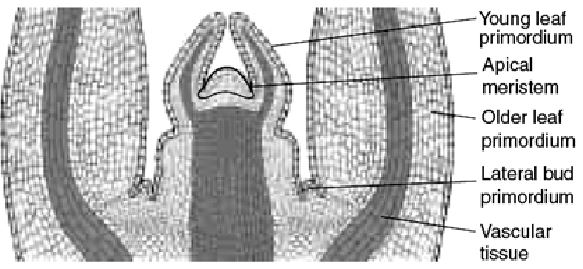
An apical shoot meristem. This longitudinal section through a shoot apex in Coleus shows the tip of a stem. Between the young leaf primordia is the apical meristem.
The meristem which occurs between mature tissues is known as intercalary meristem.
They occur in grasses and regenerate parts removed by the grazing herbivores.
Both apical meristems and intercalary meristems are primary meristems because they appear early in life of a plant and contribute to the formation of the primary plant body.
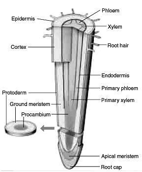
An apical root meristem. This diagram of meristems in the root shows their relation to the root tip.
The meristem that occurs in the mature regions of roots and shoots of many plants, particularly those that produce woody axis and appear later than primary meristem is called the secondary or lateral meristem.
They are cylindrical meristems. Fascicular vascular cambium, interfascicular cambium and cork-cambium are examples of lateral meristems. These are responsible for producing the secondary tissues.
Following divisions of cells in both primary and as well as secondary meristems, the newly formed cells become structurally and functionally specialised and lose the ability to divide. Such cells are termed permanent or mature cells and constitute the permanent tissues.
During the formation of the primary plant body, specific regions of the apical meristem produce dermal tissues, ground tissues and vascular tissues.
PERMANENT TISSUES
A permanent tissue is group of cells which are differentiated and which have lost the power of cell division.
The permanent tissues are composed of mature cells. The cells assume a definite shape, size and function. They lose the power of cell division.
They develop from the meristems.
Permanent tissues having all cells similar in structure and function are called simple tissues.
Permanent tissues having many different types of cells are called complex tissues.
SIMPLE TISSUES
A simple tissue is made of only one type of cells.
The various simple tissues in plants are parenchyma, collenchyma and sclerenchyma.
Parenchyma (Grew,1682)
Parenchyma is the most common simple tissue occurring in softer parts of the plant body like epidermis, cortex, pericycle, medulla, and mesophyll, pulp of fleshy fruits, embryo and endosperm.
It forms the ground tissue in which other tissues mainly vascular tissues are embedded.
It is the most primitive tissue. From this tissue other tissues have been evolved. Hence it is called the fundamental tissue.
Parenchyma forms the major component within organs. The cells of the parenchyma are generally isodiametric.
They may be spherical, oval, round, polygonal or elongated in shape. Their walls are thin and made up of cellulose.
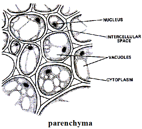
They may either be closely packed or have small intercellular spaces.
The parenchyma performs various functions like photosynthesis, storage, secretion.
The parenchyma cells of aquatic plants have fairly large intercellular spaces filled with air. They provide aeration and buoyancy to the aquatic plants.
The parenchyma cells of succulent xerophytes store water. The cells are large, vaculoes are prominent. Cell walls are thin.
The parenchyma cells of epidermis have cutinised cell walls. They are protective in function. They prevent the evaporation of water.
The parenchyma cells occurring in xylem help in conduction of water and food materials.
The parenchyma cells which are in turgid condition provide mechanical strength in herbaceous plants.
The parenchyma cells store waste products like tannins, gum, resins, oils, calcium oxalate crystals, etc.
The parenchyma cells can regain the power of cell division. Such cells are concerned in healing of wounds, formation of adventitious roots and buds and origin of secondary meristems.
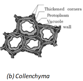
Collenchyma (Schleiden, 1839)
Collenchyma is a living, simple, mechanical tissue occurring in young stems, petioles of leaves, stalks of flowers and midribs of leaves.
It is present as a continuous band beneath the epidermis (eg. Helianthus) or in patches beneath the epidermis (eg. Cucurbita). It is absent in underground parts.
It occurs rarely in roots which are exposed to sunlight.
It is absent in leaves and stems of monocots.
It consists of cells which are much thickened at the corners due to a deposition of cellulose, hemicellulose and pectin.
Collenchymatous cells may be oval, spherical or polygonal and often contain chloroplasts.
These cells assimilate food when they contain chloroplasts.
Primary pit fields are present.
It is unevenly thickened. The thickenings are confined only to the corner of the cells.
As a result intercellular space is reduced or absent.
Some cells may contain tannins.
They provide mechanical support to the growing parts of the plant such as young stem and petiole of a leaf.
Collenchyma containing chloroplasts may carry out the process of photosynthesis.
Collenchyma may regain the power of cell division and form secondary meristems.
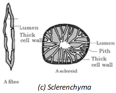
Sclerenchyma (Mettenius, 1805)
Sclerenchyma consists of long, narrow cells with thick and lignified cell walls having a few or numerous pits.
They are usually dead and without protoplasts.
On the basis of variation in form, structure, origin and development, sclerenchyma may be either fibres or sclereids.
The fibres are thick-walled, elongated and pointed cells, generally occurring in groups, in various parts of the plant.
The sclereids are spherical, oval or cylindrical, highly thickened dead cells with very narrow cavities (lumen).
These are commonly found in the fruit walls of nuts; pulp of fruits like guava, pear and sapota; seed coats of legumes and leaves of tea.
Sclerenchyma provides mechanical support to organs.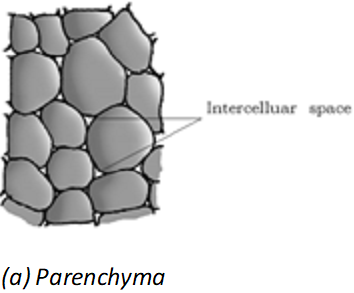
COMPLEX TISSUES
A group of different types of cells which act as a functional unit and perform a common function constitutes a complex tissue.
In vascular plants xylem and phloem are the complex tissues.
XYLEM
The term ‘Xylem” is coined by Nageli.
Xylem functions as a conducting tissue for water and minerals from roots to the stem and leaves.
It also provides mechanical strength to the plant parts.
It is composed of four different kinds of elements, namely, tracheids, vessels, xylem fibres and xylem parenchyma.
Tracheids
Tracheids are primitive vascular elements.
Tracheids are elongated or tube like cells with thick and lignified walls and tapering ends.
Tracheids having a large lumen as compared to the fibres
Tracheids join together form their ends to form a long rows. These rows extending from the roots via stem to the leaves.
Tracheids are dead and lignified cells. The deposition of lignin on cell wall is responsible to form a different type of thickenings. Pits are unlignified areas on lignified wall.
In flowering plants, tracheids and vessels are the main water transporting elements.
Bordered pits are mainly present in the wall of tracheids.
The maximum bordered pits are found in the tracheids of Gymnosperm plants.
Maximum deposition of lignin in pitted thickening.
Annular and Spiral thickening of lignin is found in protoxylem.
Reticulate and Pitted thickening of lignin is found in metaxylem.
Tracheids of pteridophytes have long or elongated bordered pits. Such type of pits is called scalariform pits.
Vessels
It is advance conductive element of xylem. The basic structure of vessels is the same as tracheids.
Vessel is a long cylindrical tube-like structure made up of many cells called vessel members, each with lignified walls and a large central cavity.
The vessel cells are also devoid of protoplasm.
Vessel members are interconnected through perforations in their common walls.
The presence of vessels is a characteristic feature of angiosperms.
Note worthy Points
1. Vessels are only found in xylem of angiosperm but exceptionally it is also present in some Gymnosperms like Ephedra, Gnetum and Welwitschia.
2. Vessels are absent in some Angiospermic plants such as Dracaena, Yucca, Dazinaria, Drimys. There are some angiosperm families in which vessel less angiosperms are included. e.g. Vinteraceae, tetracentraceae and trochodendraceae.
3. Vessels are example of dead syncyte.
Xylem Fibres
This is also dead part of xylem.
Xylem fibres have highly thickened walls and obliterated central lumens.
Xylem fibres provide strength to the tracheids and vessels.
These may either be septate or aseptate.
Xylem Parenchyma
The parenchyma found in the xylem is called xylem parenchyma. It is also called wood parenchyma.
Xylem parenchyma cells are living and thin-walled, and their cell walls are made up of cellulose.
They store food materials in the form of starch or fat, and other substances like tannins.
The radial conduction of water takes place by the ray parenchymatous cells.
They are found in primary xylem and secondary xylem.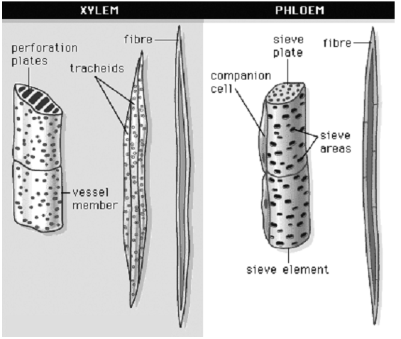
Primary xylem is of two types – protoxylem and metaxylem.
The first formed primary xylem elements are called protoxylem and the later formed primary xylem is called metaxylem.
In stems, the protoxylem lies towards the centre (pith) and the metaxylem lies towards the periphery of the organ. This type of primary xylem is called endarch.
In roots, the protoxylem lies towards periphery and metaxylem lies towards the centre. Such arrangement of primary xylem is called exarch.
PHLOEM
The term ‘Phloem’ is coined by Nageli (1858).
The main function of phloem is transportation of organic materials from leaves to other parts of the plant.
Phloem is a heterogeneous tissue.
Phloem in angiosperms is composed of four types of cells. They are sieve tube elements, companion cells, phloem parenchyma and phloem fibres.
Gymnosperms have albuminous cells and sieve cells. They lack sieve tubes and companion cells.
Sieve Tube Elements
Sieve element is discovered by Harting.
Sieve tube elements are also long, tube-like structures, arranged longitudinally and are associated with the companion cells.
Their end walls are perforated in a sieve-like manner to form the sieve plates.
A mature sieve element possesses a peripheral cytoplasm and a large vacuole but lacks a nucleus.
The functions of sieve tubes are controlled by the nucleus of companion cells.
Companion cells
Companion cells are special parenchyma cells associated with sieve tubes.
Usually one companion cell associates with a sieve tube.
Occasionally more than one companion cell arises from the same mother cell by a longitudinal division.
It consists of thin cellulose and vacuolated cytoplasm and a big nucleus. It shows cytoplasm connections with sieve tube through plasmodesmata.
Companion cell is found only in angiosperms.
They are absent in pteridophytes and gymnosperms where albuminous cells are associated with sieve cells.
Companion cells assist sieve tubes in translocation of food materials.
Phloem Parenchyma
Parenchyma present in phloem is called phloem parenchyma.
The cells of phloem parenchyma may be longer, broader, polyhedral or sub spherical in shape.
The cells are living and surrounded by thin cellulose cell walls.
Cell walls show primary pits fields.
Primary phloem consists of vertically elongated phloem parenchyma. Such phloem is called axial phloem parenchyma.
Secondary phloem consists of axial and ray parenchyma.
Axial parenchyma consists of vertically elongated cells.
Ray parenchyma consists of radial elongated cells.
Phloem parenchyma is found in dicots and some monocots. It is absent in many monocots.
Phloem parenchyma helps in storage of food materials and in radial conduction of materials.
Phloem Fibers
The fibers present in phloem are called phloem fibers. They are also called as bast fibres. They are long and narrow with pointed ends. They overlap with each other.
These are generally absent in the primary phloem but are found in the secondary phloem.
The fibers of secondary phloem have lignified walls.
The walls show simple pits. Rarely bordered pits may be present. The fibers may be living or dead.
Living fibers store food materials.
At maturity, these fibres lose their protoplasm and become dead. Dead fibers provide rigidity and mechanical support to the plant body.
Phloem fibres of jute, flax and hemp are used commercially.
The first formed primary phloem consists of narrow sieve tubes and is referred to as protophloem and the later formed phloem has bigger sieve tubes and is referred to as metaphloem.
2.2 The Tissue System
We were discussing types of tissues based on the types of cells present. Let us now consider how tissues vary depending on their location in the plant body. Their structure and function would also be dependent on location. On the basis of their structure and location, there are three types of tissue systems. These are the epidermal tissue system, the ground or fundamental tissue system and the vascular or conducting tissue system.
EPIDERMAL TISSUE SYSTEM
The epidermal tissue system forms the outer-most covering of the whole plant body and comprises epidermal cells, stomata and the epidermal appendages – the trichomes and hairs.
Epidermis
The primary regions of the plant body are covered by a layer of epidermal tissue system. This layer is called epidermis.
It is made up of elongated, compactly arranged cells, which form a continuous layer without any intercellular spaces. Epidermis is usually single- layered.
In surface view the cells are isodiametric in shape. In transverse section they are rectangular in shape.
In monocotyledonous stems and leaves they are elongated in the direction of long axis.
Epidermal cells are parenchymatous with a small amount of cytoplasm lining the cell wall and a large vacuole.
The outside of the epidermis is often covered with a waxy thick layer called the cuticle which prevents the loss of water.
Cuticle is absent in roots.
Chloroplasts are absent. They are present in epidermal cells of aquatic plants and plants growing in moist and shady places. Epidermal cells retain the power of division.
Bulliform Cells
They are large, thin walled and highly vacuolated cells found in the epidermis of leaves of many monocots. The walls of these cells are usually thin. The outer walls may be thick. The cells mainly contain water. Plastids are absent. The cells may be present on both sides of a leaf, but they are more common on the upper side.
Bulliform cells are concerned with the unrolling of developing leaves. They play a role in hygroscopic opening and closing of mature leaves due to changes in turgor pressure. They are also concerned with water-storage.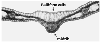
Stomata
Stomata are the minute pores present in the epidermis of aerial parts of the plant body.
Each stoma is composed of two bean- shaped cells known as guard cells.
In grasses, the guard cells are dumb- bell shaped.
The outer walls of guard cells (away from the stomatal pore) are thin and the inner walls (towards the stomatal pore) are highly thickened.
The guard cells possess chloroplasts and regulate the opening and closing of stomata.
The epidermal cells surrounding the guard cells differ from the other epidermal cells in size and arrangement. These epidermal cells are called subsidiary or accessory cells.
The stomatal aperture, guard cells and the surrounding subsidiary cells are together called stomatal apparatus or stomatal complex.
In dorsiventral leaves they are present on lower surface.
In isobilateral leaves they are present on the both surfaces.
In floating leaves they are present on upper surface.
Stomata present on the floral parts and in aquatic plants are functionless.
Stomata are absent in roots and woody stems.
Stomata regulate the process of transpiration and gaseous exchange.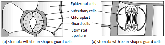
Epidermal appendages
The cells of epidermis bear a number of hairs.
The root hairs are unicellular elongations of the epidermal cells and help absorb water and minerals from the soil.
On the stem the epidermal hairs are called trichomes.
The trichomes in the shoot system are usually multicellular.
They may be branched or unbranched and soft or stiff. They may even be secretory.
The trichomes help in preventing water loss due to transpiration.
THE GROUND TISSUE SYSTEM
Ground tissues system includes all the tissues except epidermal and vascular tissue system.
It is largest and exhaustive tissue system.
It occupies larger areas of the plant body.
It begins from the layer just beneath the epidermis and continues up to the centre of the plant body.
It mainly consists of parenchyma. Collenchyma, sclerenchyma, laticiferous tissue secretory tissue, etc, may be present.
Parenchymatous cells are usually present in cortex, pericycle, pith and medullary rays, in the primary stems and roots.
In leaves, the ground tissue consists of thin-walled chloroplast containing cells and is called mesophyll.
THE VASCULAR TISSUE SYSTEM
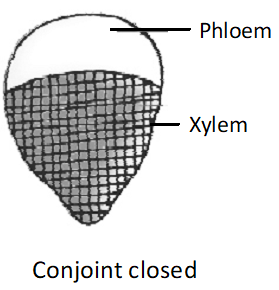
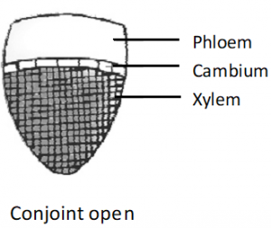
The vascular tissue comprises xylem and phloem. The vascular bundles may be radial, conjoint and concentric.
Radial vascular bundles found in roots, conjoint vascular bundles found in stem.
The conjoint bundle may be collateral and bicollateral.
The concentric bundles may be amphivasal (leptocentric) or amphicribal (hedrocentric).
On the basis of cambium, the bundles may be open (dicot stem) or closed (monocot stem). On the basis of relative position of protoxylem and metaxylem, the xylem may be exarch, endarch or mesarch.
Depending upon the number of protoxylem elements, the root may be mono-, di-, tri-, tetra-, penta-, hexa- or polyarch.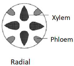
Various types of vascular bundles
ANIMAL TISSUES
2.3 INTRODUCTION
When most people think of animals, they think of their pet dogs and cats and the animals that they have seen in a zoo, on a farm, in an aquarium, or out in the wild. When they think about the diversity of animals, they may think of the differences between the predatory lions and tigers and the herbivorous deer and antelope, between a ferocious- looking shark and a playful dolphin. Despite the differences among these animals, they are all vertebrates. All vertebrates share the same basic body plan, with the same sorts of organs operating in much the same way.
The human body is composed of billions of cells to perform various functions. How do these cells in the body work together? In multicellular animals, a group of similar cells along with intercellular substances perform a specific function. Such an organisation is called tissue. All complex animals consist of only four basic types of tissues. These tissues are organized in specific proportion and pattern to form an organ like stomach, lung, heart and kidney. When two or more organs perform a common function by their physical and/or chemical interaction, they together form organ system, e.g., digestive system, respiratory system, etc. Cells, tissues, organs and organ systems split up the work in a way that exhibits division of labour and contribute to the survival of the body as a whole.
2.4 ANIMAL TISSUES
Based on structure tissues are different and are broadly classified into four types: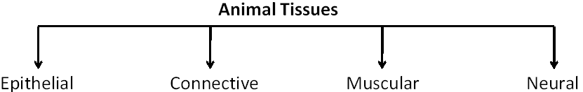
EPITHELIAL TISSUE
Epithelial tissues provide covering to the inner and outer lining of various organs. The cells of epithelial tissues are compactly packed with little intercellular matrix.
There are two types of epithelial tissues:
(a) Simple Epithelium: Simple epithelium is composed of a single layer of cells and functions as a lining for body cavities, ducts, and tubes.
(b) Compound Epithelium: The compound epithelium consists of two or more cell layers and has protective function as it does in our skin. They cover the dry surface of the skin, the moist surface of buccal cavity, pharynx, inner lining of ducts of salivary glands and of pancreatic ducts.
On the basis of structural modification of the cells, simple epithelium is further divided into three types. These are: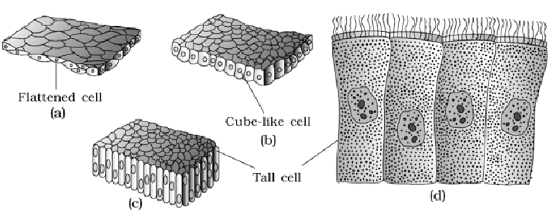
Simple epithelium: (a) Squamous (b) Cuboidal (c) Columnar(d) Columnar cells bearing cilia
(a) Squamous: The squamous epithelium is made of a single thin layer of flattened cells with irregular boundaries. They are found in the walls of blood vessels and air sacs of lungs and are involved in functions like forming a diffusion boundary.
(b) Cuboidal: The cuboidal epithelium is composed of a single layer of cube-like cells. This is commonly found in ducts of glands and tubular parts of nephrons in kidneys and its main functions are secretion and absorption. The epithelium of proximal convoluted tubule (PCT) of nephron in the kidney has microvilli.
(c) Columnar: The columnar epithelium is composed of a single layer of tall and slender cells. Their nuclei are located at the base. Free surface may have microvilli. They are found in the lining of stomach and intestine and help in secretion and absorption. If the columnar or cuboidal cells bear cilia on their free surface they are called ciliated epithelium. Their function is to move particles or mucus in a specific direction over the epithelium. They are mainly present in the inner surface of hollow organs like bronchioles and fallopian tubes.
Some of the columnar or cuboidal cells get specialised for secretion and are called glandular epithelium. They are mainly of two types: unicellular, consisting of isolated glandular cells (goblet cells of the alimentary canal), and multicellular, consisting of cluster of cells (salivary gland). On the basis of the mode of pouring of their secretions, glands are divided into two categories namely exocrine and endocrine glands. 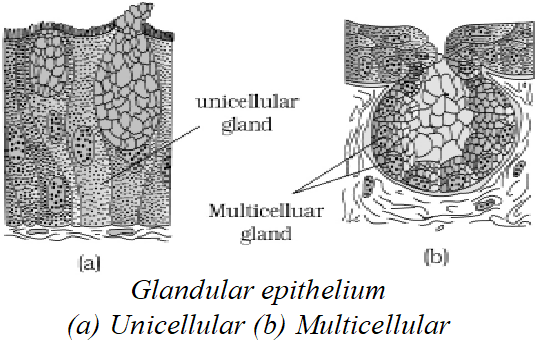
Exocrine glands secrete mucus, saliva, earwax, oil, milk, digestive enzymes and other cell products. These products are released through ducts or tubes. In contrast, endocrine glands do not have ducts. Their products called hormones are secreted directly into the fluid bathing the gland.
Compound epithelium is made of more than one layer (multi-layered) of cells and thus has a limited role in secretion and absorption. Their main function is to provide protection against chemical and mechanical stresses. They cover the dry surface of the skin, the moist surface of buccal cavity, pharynx, inner lining of ducts of salivary glands and of pancreatic ducts. All cells in epithelium are held together with little intercellular material. In nearly all animal tissues, specialised junctions provide both structural and functional links between its individual cells.
JUNCTIONS
Three types of cell junctions are found in the epithelium and other tissues. These are called as tight, adhering and gap junctions. Tight junctions help to stop substances from leaking across a tissue. Adhering junctions perform cementing to keep neighbouring cells together. Gap junctions facilitate the cells to communicate with each other by connecting the cytoplasm of adjoining cells, for rapid transfer of ions, small molecules and sometimes big molecules.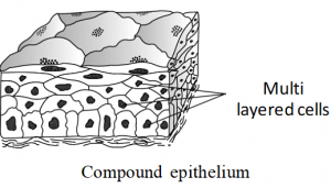
Summary of Epithelial Tissues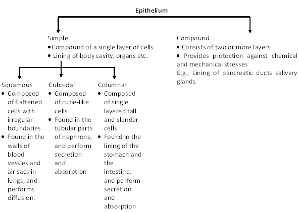
CONNECTIVE TISSUE
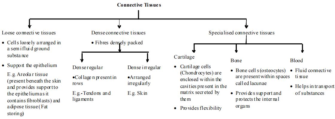
Connective tissues are most abundant and widely distributed in the body of complex animals. Function of connective tissues is linking and supporting other tissues/organs of the body. In all connective tissues except blood, the cells secrete fibres of structural proteins called collagen or elastin. The fibres provide strength, elasticity and flexibility to the tissue. These cells also secrete modified polysaccharides, which accumulate between cells and fibres and act as matrix (ground substance).
Connective tissues are classified into three types:
1. Loose Connective Tissue: Loose connective tissue has cells and fibres loosely arranged in a semi-fluid ground substance, for example, areolar tissue present beneath the skin. Often it serves as a support framework for epithelium. It contains fibroblasts (cells that produce and secrete fibres), macrophages and mast cells. Adipose tissue is another type of loose connective tissue located mainly beneath the skin. The cells of this tissue are specialised to store fats. The excess of nutrients which are not used immediately are converted into fats and are stored in this tissue.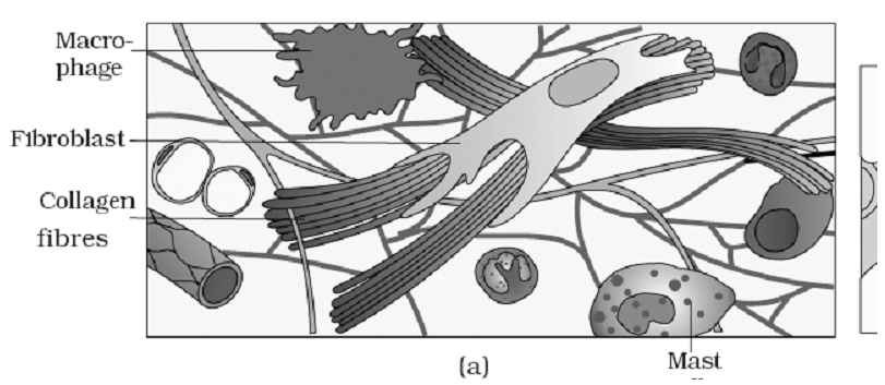
Loose connective tissue : (a) Areolar tissue (b) Adipose tissue
2. Dense Connective Tissue: Fibres and fibroblasts are compactly packed in the dense connective tissues. Orientation of fibres show a regular or irregular pattern and are called dense regular and dense irregular tissues. In the dense regular connective tissues, the collagen fibres are present in rows between many parallel bundles of fibres. Tendons, which attach skeletal muscles to bones and ligaments which attach one bone to another are examples of this tissue. Dense irregular connective tissue has fibroblasts and many fibres (mostly collagen) that are oriented differently. This tissue is present in the skin.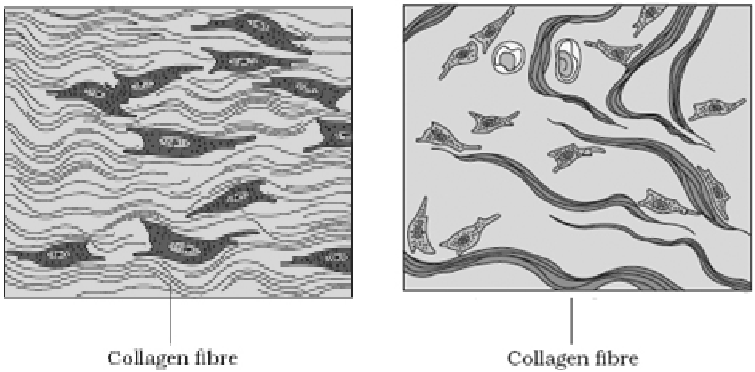
Dense connective tissue: (a) Dense regular (b) Dense irregular
3. Specialised Connective Tissue: Cartilage, bones and blood are various types of specialized connective tissues.
Cartilage: The intercellular material of cartilage is solid and pliable and resists compression. Cells of this tissue (chondrocytes) are enclosed in small cavities within the matrix secreted by them. Most of the cartilages in vertebrate embryos are replaced by bones in adults. Cartilage is present in the tip of nose, outer ear joints, between adjacent bones of the vertebral column, limbs and hands in adults.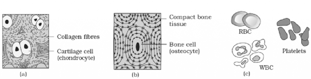
Bones: Bones have a hard and non-pliable ground substance rich in calcium salts and collagen fibres which give bone its strength. It is the main tissue that provides structural frame to the body. Bones support and protect softer tissues and organs. The bone cells (osteocytes) are present in the spaces called lacunae. Limb bones, such as the long bones of the legs, serve weight-bearing functions. They also interact with skeletal muscles attached to them to bring about movements. The bone marrow in some bones is the site of production of blood cells.
Blood: Blood is a fluid connective tissue containing plasma, red blood cells (RBC), white blood cells (WBC) and platelets. It is the main circulating fluid that helps in the transport of various substances.
Summary of Connective Tissues
MUSCLE TISSUE
Muscle: Each muscle is made of many long, cylindrical fibres arranged in parallel arrays. These fibres are composed of numerous fine fibrils, called myofibrils. Muscle fibres contract (shorten) in response to stimulation, then relax (lengthen) and return to their uncontracted state in a coordinated fashion. Their action moves the body to adjust to the changes in the environment and to maintain the positions of the various parts of the body. In general, muscles play an active role in all the movements of the body.
TYPES OF MUSCLES
Muscles are of three types:
Skeletal Muscle: Skeletal muscle tissue is closely attached to skeletal bones. In a typical muscle such as the biceps, striated (striped) skeletal muscle fibres are bundled together in a parallel fashion. A sheath of tough connective tissue encloses several bundles of muscle fibres.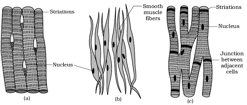
Figure: Muscle tissue : (a) Skeletal muscle tissue (b) Smooth muscle tissue (c) Cardiac muscle tissue
Smooth Muscle: The smooth muscle fibres taper at both ends (fusiform) and do not show striations. Cell junctions hold them together and they are bundled together in a connective tissue sheath. The wall of internal organs such as the blood vessels, stomach and intestine contains this type of muscle tissue. Smooth muscles are ‘involuntary’ as their functioning cannot be directly controlled.
Cardiac Muscle: Cardiac muscle tissue is a contractile tissue present only in the heart. Cell junctions fuse the plasma membranes of cardiac muscle cells and make them stick together. Communication junctions (intercalated discs) at some fusion points allow the cells to contract as a unit, i.e., when one cell receives a signal to contract, its neighbours are also stimulated to contract.
|
Skeletal muscle fibres |
Smooth muscle fibres |
Cardiac muscle fibres |
|
1. They are very long and cylindrical with blunt ends. 2. Multinucleate, nuclei just near the sarcolemma. 3. Fibres are unbranched. 4. Voluntary. 5. No intercalated discs. 6. Mitochondria are moderately abundant. 7. They contract quickly.
|
1. They are long and spindle-shaped with tapering ends. 2 Uninucleate, nucleus at the centre. 3. Fibres are unbranched. 4. Involuntary. 5. No intercalated discs. 6. Mitochondria are fewer.
|
1. Short and cylindrical with truncate ends. 2. Mostly uninucleate, nucl eus at the centre. 3. Fibres are branched. 4. Involuntary. 5. Intercalated discs occur between the ends of fibres. 7. They show rhythmic contractions. 8. Also called heart muscle fibres. |
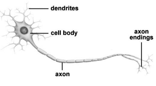
NEURAL TISSUE
Neurons, the unit of neural system are excitable cells. The neuroglial cell which constitute the rest of the neural system protect and support neurons. Neuralgia make up more than one-half the volume of neural tissue in our body. When a neuron is suitably stimulated, an electrical disturbance is generated which swiftly travels along its plasma membrane. Arrival of the disturbance at the neuron’s endings, or output zone, triggers events that may cause stimulation or inhibition of adjacent neurons and other cells and thereby transmitting neural signals to different parts of the body.