


1. CELL CYCLE AND CELL DIVISION
Growth and reproduction are characteristics of cells, indeed of all living organisms. All cells reproduce by dividing into two, with each parental cell giving rise to two daughter cells each time they divide. These newly formed daughter cells can themselves grow and divide, giving rise to a new cell population that is formed by the growth and division of a single parental cell and its progeny. In other words, such cycles of growth and division allow a single cell to form a structure consisting of millions of cells.
The process of formation of daughter cells from the parent cell is known as cell division, cell multiplication or cell reproduction. The cell reproduction is one of the fascinating characteristics of living organisms. Boveri and Flemming (1879) studied the process of formation of two identical cells from the preexisting cell. The daughter cells through half the size of the parent cell have all the potentialities of the parent cell to survive, grow and reproduce.
There are two types of cell division seen in eukaryotic organisms. They are mitosis and meiosis. Mitosis is the process of producing two identical daughter cells by a parent cell, each daughter cell has same amount of genetic material (same number of chromosomes) as the parent cell. Hence mitosis is known as replicative division. Mitosis is usually seen in the somatic cells of higher plants and animals, whereas in unicellular organisms it is a means of asexual reproduction producing many progeny.
Meiosis is the process of producing four daughter cells by a parent cell, where in each daughter cell has half the amount of genetic material (half the number of chromosomes) as the parent cell. It is also known as reductive division since it reduces the number of chromosomes in the daughter cells to half the number of chromosomes of the parent cell. The meiosis occurs in the gamete forming tissues or cells of the sexually reproducing organisms.
2. CONCEPT OF CELL CYCLE
Cell Cycle
Cell division is a very important process in all living organisms. During the division of a cell, DNA replication and cell growth also take place. All these processes, i.e., cell division, DNA replication, and cell growth, hence, must take place in a coordinated way to ensure correct division and formation of progeny cells containing intact genomes. The sequence of events by which a cell duplicates its genome, synthesizes the other constituents of the cell and eventually divides into two daughter cells is termed cell cycle. Although cell growth (in terms of cytoplasmic increase) is a continuous process, DNA synthesis occurs only during one specific stage in the cell cycle. The replicated chromosomes (DNA) are then distributed to daughter nuclei by a complex series of events during cell division. These events are themselves under genetic control.
The time period required to complete one cell cycle i.e. from the beginning of one cell division to the beginning of next is called generation time. It varies from 30 minutes in bacteria to 90 minutes, in yeast. Though the details of cell cycle vary from organism to organism, certain events are universal.
Phases of Cell Cycle
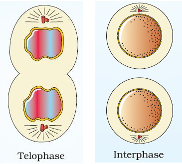
A typical eukaryotic cell cycle is illustrated by human cells in culture. These cells divide once in approximately every 24 hours. However, this duration of cell cycle can vary from organism to organism and from cell type to cell type. Yeast for example, can progress through the cell cycle in only about 90 minutes. The cell cycle is divided into two basic phases: i.e., Interphase and M-phase
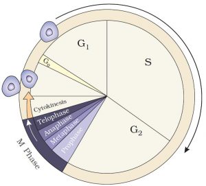
Interphase (L., inter = between; phases = stage)
This is the period between two successive mitotic divisions. It is a period of active synthesis and intense growth. During this stage the genes are active, and the cell synthesizes most of its components to double its size. In a typical animal cell one cell cycle may take 24 hours to complete. The mitosis usually takes 1 to 2 hours and the inter-phase lasts for 22 to 23 hours, i.e., more than 95% of total cell-cycle.
The inter-phase can be divided into three phases. They are G1 (gap 1) phase, S (synthesis) phase and G1 (gap 2).
G1 phase (Gap I, First Growth phase post mitotic, pre-synthetic phase)
G1 phase occupies a major portion of cell cycle. In a typical animal cell, it lasts for about 10 hours. It is a period of intensive synthesis of cellular components (except DNA). The cell organelles such as mitochondria, chloroplast, lysosomes, ER, Golgi apparatus are produced. The synthesis of tRNA, rRNA, mRNA and ribosomes occur. The metabolic rate of the cell will be very high. The cell during the G1 phase produces structural and functional proteins. The cell may get prepared to get into S phase. During the G1 phase depending upon the conditions the substances are produced that may stimulate or inhibit the onset of S phase.
To proceed or not to proceed to the next phase of cell cycle is controlled by regulatory substances called protein kinases. These for their activity depend on proteins called cyclins, hence are known as cyclin-dependent protein kinases (CDKs). There are different types of cyclins for the different stages of cell cycle. Each of them is synthesized at the required stage and destroyed at the end of the specific stage. For initiating G1 phase G1 specific cyclins are synthesized and they activate CDKs specific for G1 phase.
The G1 specific cyclins are destroyed at the end of G1 phase and S phase specific cyclin are synthesized. This initiates S phase.
S phase (Synthetic phase).
During S phase DNA replication occurs. Proteins associated with the chromosomes, namely histones are synthesized. The chromosomes are duplicated, to form two longitudinal halves known as two chromatids attached to each other at a centromere to form a dyad. The DNA content is doubled from 1c to 2c in a haploid cell and 2c to 4c in a diploid cell (The quantity of DNA present in a haploid cell is 1c). Hence this phase is known as Invisible M phase.
In animal cells, during the S phase, DNA replication begins in the nucleus, and the centriole duplicates in the cytoplasm.
At the end of S phase S specific cyclins are destroyed. The S phase lasts for 8-9 hours. During S and G2 phases the mitotic related cyclins are synthesized. They accumulate in the cell and combine with specific CDKs. These M (Mitosis) phase CDKs are kept in an inactive state till G2 phase is completed and the DNA has been completely duplicated and undamaged.
G2 phase
The gap-2 phase is characterized by the second phase of intense cellular synthesis. The multiplication of chloroplasts and mitochondria occurs. The centrioles divide, the proteins of spindle fibres are synthesized and spindle begins to form. The protein of the spindle fibres is called tubulin and it polymerizes to form spindle fibres. There will be active metabolism leading to the storage of energy for the operation of M phase. The G2 phase usually lasts for 5-6 hours.
G0 phase
It is a quiescent stage in which a cell does not divide further and escapes from cell cycle in early G1 and differentiates to perform specific functions. E.g., nerve cell skeletal muscles cell, matured WBCs, RBCs etc. Some cells on G0 are metabolically active and if stimulated return to cell cycle through differentiation and complete divisions. E.g., fibroblasts to heal wounds, parenchyma cells in plants to form secondary meristems.
3. M PHASE
This is the most dramatic period of the cell cycle, involving a major reorganization of virtually all components of the cell. Since the number of chromosomes in the parent and progeny cells is the same, it is also called as equational division. Though for convenience mitosis has been divided into four stages of nuclear division, it is very essential to understand that cell division is a progressive process and very clear-cut lines cannot be drawn between various stages. Mitosis is divided into the following four stages:
- Prophase
- Metaphase
- Anaphase
- Telophase
Mitosis is the common type of cell division where the chromosomes duplicated during the interphase are equally distributed to the daughter nuclei. Mitosis involves two processes namely karyokinesis or nuclear division and cytokinesis or cytoplasmic division. The karyokinesis through a continuous process, for the sake of convenience of understanding is divided into four phases. The phases are prophase, metaphase, anaphase, and telophase.
Prophase
- Prophase is the first phase of karyokinesis usually the longest phase of mitosis. It has three substages, namely, early, middle and late prophase.
Early prophase: The chromatin fibres undergo condensation by coiling and dehydration to form short and thick chromatids. - The chromatids (the duplicated chromosomes occurs in the form of a pair of chromatids held together at the centromere) become short and thick thread like structures due to condensation of DNA and protein and also due to coiling (spiralization).
- The centrioles (in the animal cells) begin to move towards the opposite poles of the cell. The centrioles develop short microtubules radiating from them and together from asters. In plant cells centrioles and absent hence spindle is anastral.
- The centrioles with spindle fibres constitute the mitotic spindle.
Mid prophase: The nuclear envelop becomes fragmented and at the end of prophase it disappears. - The nucleoli gradually decrease in size and finally disappear. The mitotic spindle becomes established.
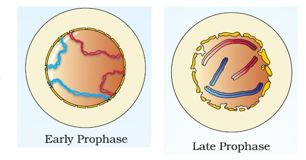
The chromosomes undergo further condensation become shorter thicker and darkly stained. The paired chromatids are attached to centromere
Late prophase or pro metaphase: Then nuclear envelope and nucleoli disappear, and chromosomes are released to the cytoplasm. The Golgi apparatus and endoplasmic reticulum become inconspicuous. The spindle apparatus is organized.
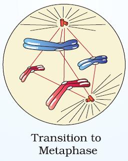
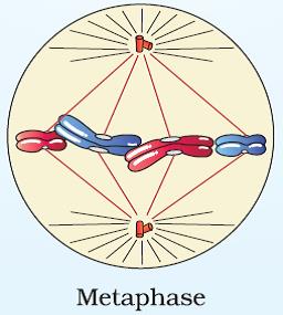
Metaphase
The complete disintegration of the nuclear envelope marks the start of the second phase of mitosis; hence the chromosomes are spread through the cytoplasm of the cell.
It is a short phase and lasts 2-10 minutes. It is known as the stage of orientation.
- The chromosomes in the form of paired chromatids become attached to the spindle by certain spindle fibres. These spindle fibres are called chromosomal, kinetochore or tactile fibres. The chromosomal fibres are connected to two C shaped or disc shaped kinetochores covering the centromere of each chromosome.
The metaphase is characterized by all the chromosomes coming to lie at the equator with one chromatid of each chromosome connected by its kinetochore to spindle fibres from one pole and its sister chromatid connected by its kinetochore to spindle fibres from the opposite pole. The plane of alignment of the chromosomes at metaphase is referred to as the metaphase plate. - The chromosomes move towards the ‘equator’ of spindle in such a manner that their centromeres are aligned along the equatorial plate which lies in the middle and at right angles to the spindle.
- The centromere of each chromosome is connected to both the asters and poles of the spindle by these chromosomal fibres.
- In the spindle there are three types of fibres. They are (a) continuous fibres or polar fibres which extend from pole to pole of the spindle, (b) the chromosomal fibres which are attached to the centromeres of the chromosomes and extend to one of the poles and (c) discontinuous or inter polar fibres which extend from a pole to variable distance but do not reach the opposite poles.
The chromosome arranges in the form of plate called metaphase plate or equatorial plate. The process of bringing the chromosomes to the equator is known as congression. The metaphase chromosomes are maximally condensed, short, and thick. They are ideal to study the chromosome number and morphology.
Anaphase
- Anaphase is a rapid stage of migration of chromosomes and is the shortest stage (2-3 minutes).
- The centromere of each chromosome splits longitudinally into two and the chromosomal fibres pull the daughter centromeres towards the opposite poles. The separation of chromosomes occurs due to dissolution of cohesion at centromere that holds sister chromatids together.
- The separated chromatids become daughter chromosomes known as monad chromosomes.
- The arms of the chromosomes follow the centromere and move towards the poles. the metacentric, sub-metacentric, acrocentric, and telocentric chromosomes appear like V, L, j and I during anaphase.
The anaphase movement of chromosomes is caused by shortening of chromosomal fibres due to disassembly of its microtubular subunits at kinetochoral end. The chromosomes during this process move pole ward due to sliding movement of kinetochoral motor proteins dyneins which transport the chromosomes along the shortening microtubules using ATPs.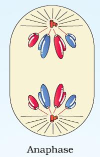
Telophase
- The telophase is of long duration.
- The chromosomes reach the poles and undergo uncoiling becoming long and slender thread like structures. They become indistinct and form the chromatin mass and lose their individuality.
- Each mass of chromatin thread becomes a daughter nucleus.
- The nuclear envelope slowly appears around the mass of chromatin from pieces of old nuclear membrane and ER and even the nucleoli reappear from Sine acido thymonucleinico (SAT) chromosome of nucleolar organizer region (NOR) region the fibrous lamina around chromatin reappear.
- In animal cells astral rays and spindle fibres and in plant cells spindle fibres near the poles disappear but they remain intact at equator to form phragmoplast (Gr., phragmos = hedge or enclosure). The Golgi bodies and ER are reformed. After telophase, the cell enters into cytokinesis.
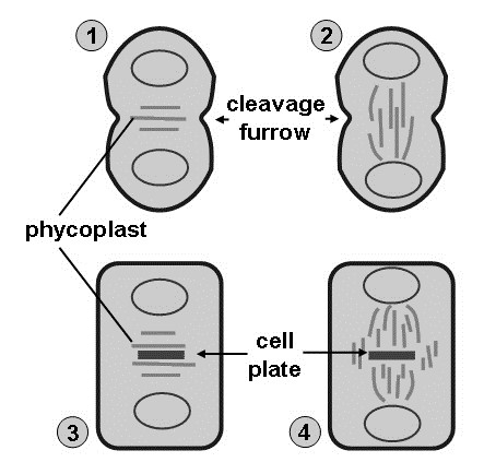
Cytokinesis
During mitosis, the organelles of the cell becomes distributed towards the two poles of the cell (The ER and Golgi apparatus break up into small fragments and become distributed towards the poles).
In animal cells cytokinesis occurs by the formation of cleavage furrow or invagination of the cell membrane at the region previously occupied by the equator of the spindle. A dense remnant of spindle material collects at the equator and is known as mid body as a bridge. The furrow is formed by a contractile band of microfilaments of actin and myosin attached to the inner surface of plasma membrane of the furrow. The constriction deepens and divides the cytoplasm into two. The mid body then disappears. The cleavage cytokinesis is also observed in some protists and pollen forming meiocytes of angiosperms. Actin filaments attached to the cell membrane contract and the furrow deepens and its membranes meet in the centre of the cell and fuse forming two complete daughter cells by centripetal cleavage.
In plant cells the spindle fibres present at the equator move outwards and increase in number forming a barrel shaped structure called phragmoplast to trap Golgi vesicles to form a cell plate. The rest of fibres of the spindle disintegrated. The vesicles from the Golgi apparatus line up along the region right angles to spindle apparatus. These vesicles fuse to form a cell plate. The cell plate extends towards the periphery along the equatorial line and meets the parent plasma membrane and fuses with it. The contents of the vesicles form the middle lamella. The membrane of the cell plate forms the new plasma membranes of the opposing daughter cells at the region of their contact. Cellulose cell walls are laid down on these new membranes. Thus, two separate daughter cells are formed. In some areas the vesicles of the cell plate do not fuse and maintain cytoplasmic contacts between daughter cells. These contacts are known as plasmodesmata. On either side of middle lamella, the cytoplasm deposits a primary cell wall of cellulose, hemicellulose and pectin thus dividing the parent cell into two daughter cells. Thus, in plant cells cytokinesis progresses centrifugally.
4. SIGNIFICANCE OF MITOSIS
- Mitosis helps in the maintenance of genetic stability. The chromosomal number is maintained constant among the daughter cells. The daughter cells are genetically identical to their parent cells.
- Mitosis is responsible for the growth and development of the multicellular organisms. The increase in tissue mass occurs due to increase in the number of cells. This process is called hyperplasia.
- Mitosis helps in maintenance of the nuclear cytoplasmic ratio. An overgrown cell is induced to divide.
- Mitosis helps in replacing worm out cells and tissues. Injured and dead tissues are replaced by the healthy cells of the specific tissue through mitosis.
- Regeneration of damaged part of an organ occurs through mitosis.
- Asexual reproduction in unicellular organism involves mitosis.
5. MEIOSIS
Meiosis is a type of cell division where the diploid number of chromosomes is reduced to haploid number. This reduction division is responsible for the maintenance of constancy of chromosome number in the sexually reproducing organisms. It is characterized by a single duplication of chromosomes in the parent cell followed by two nuclear divisions (karyokinesis) and two cell divisions (cytokinesis).
Meiosis was discovered by Edward Strasburger (1888) and the term meiosis was coined by Farmer and Moore (1905). Meiosis consists of two divisions, namely, Meiosis I and Meiosis II. There is a single interphase before meiosis I.
Meiosis I is also known as reduction division or heterotypic division. During this division, the chromosome number is reduced to half. Prior to meiosis I there is interphase where the duplicated chromosomes occur as chromatids. After interphase the chromosome number is 2n in a diploid cell, but chromatid number is double and DNA content is 4c. Each chromosome exists as a pair of chromatids held together by the centromere.
Occurrence: Meiosis occurs in specific diploid cells at a particular time. They occur in gonads of animal microspore and megaspore mother cells of plants. The cells undergoing meiosis are called meiocytes e.g., spermatocytes forming sperms. Oocytes forming ova in animals. In the sporangia of plants, the meiocytes are microspore mother cells forming microspores and megaspore mother cells forming megaspores.
Meiosis I
Meiosis I consist of karyokinesis and cytokinesis. The karyokinesis shows four phases. They are prophase I, metaphase I, anaphase I and telophase I.
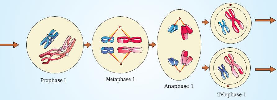
Prophase I
This is the complex and longest phase. It is divided into five sub stages namely leptotene, zygotene, pachytene, diplotene and diakinesis.
(i) Leptotene (Gr., leptos = thin; tainia = band; nema = thread)
- The chromosomes undergo condensation, spiralization and become visible.
- Even through the chromosomes have undergone duplication, the chromatids remain invisible.
- The centrioles develop asters begin to move apart towards the opposite poles.
- Bead like structures may be seen all along the length of the chromosomes. These are highly coiled regions of chromosomes.
- In many animals calls the chromosomes are converged towards the centrosomal ends to form a ‘bouquet’ stage. The ends of chromosomes are attached to nuclear envelope by a structure called attachment plate.
(ii) Zygotene (Gr., zygos = yoke, pair; tainia = band; nema = thread)
- The homologous chromosomes undergo pairing. The process is called ‘synapsis’. There are three types of synapsis – pro centric starting from the centromere, proterminal starting from the tips and random starting at many points.
- The pairing occurs in a “zipper” like fashion. The pairing of homologous chromosomes is due to the formation of ladder like protein complex called the synaptonemal complex. This aligns the homologous chromosomes accurately placing the corresponding genetic regions of chromosome pair opposite to each other like a scaffold.
- The paired homologous chromosomes are called bivalents.
- The chromatids of the chromosomes are still invisible.
(iii) Pachytene (Gr., pachus = thick)
- The first two stages of prophase I are relatively short-lived compared to the next stage that is pachytene.
- Pairing is completed.
- The chromosomes become further thick and short.
- The two chromatids of a chromosome are called sister chromatids or dyads and the chromatids of the two members of the homologous chromosomes are called non-sister chromatids tetrads.
- Crossing over is also an enzyme-mediated process and the enzyme involved is called recombinase.
- The phenomenon of exchange of genetic material occurs between the non-sister chromatids of each homologous pair of chromosomes. This occurs by breakage called nicking, by endonuclease and reunion is called annealing. This is helped by ligase. These enzyme completes together known as recombination nodules. This is known as crossing over and it results in the recombination of genes in the chromosomes.
- The bivalents are composed of four chromatids, hence are called tetrads. The tetrad nature is not yet visible.
(iv) Diplotene (Gr., diplos = double)
- The synaptonemal complex formed between the homologous chromosomes begin to dissolve.
- The homologous chromosomes repel each other and start separating this is known as repulsion. The homologous chromosomes are attached at certain ‘X’ shaped region called chiasmata. The diplotene stage may be prolonged and arrested for 8-10 years as in human female from 5th month of foetal stage up to 12th year during the start of ovulation. In the oocytes of vertebrates – fishes, amphibians, reptiles, and birds the diplotene chromosomes form giant lamp brush chromosomes with lateral extended loops helping in transcription of m-RNA, r-RNA to synthesize proteins of yolk.
- The tetrad becomes visible.
- The regions of crossing over where exchange of genetic material occurs between the non-sister chromatids of the homologous chromosomes appear as ‘X’ like structures called chiasmata.
(Chiasmata are cytological demonstration of the genetic phenomenon of crossing over).
(v) Diakinesis (Gr., dia = across; kinesis = movement)
- The chromosomes become very thick and short
- The repelling of bivalents continues
- The chiasmata slide towards the tips disappear one after another by moving towards the tips of the chromatids. This process is called terminalization.
- The nuclear membrane and nucleolus disappear and release the tetrads freely in the cytoplasm.
- Spindle fibres begin to form.

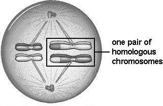
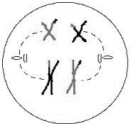
Metaphase I
- The tetrads become attached to the spindle fibres through their kinetochores (Centromeres).
- The kinetochores of maternal and paternal chromosomes (the two members of each homologous pair of chromosomes) face opposite spindle poles and get connected to the facing pole through chromosomal fibres randomly.
- The tetrads move to the equator of the spindle.
- On either side of the equator of the spindle there is a haploid set of chromosomes forming two metaphase plates of n’ set of chromosomes thus two parallel metaphase plates are formed. Each set of chromosomes is connected to only one pole of the spindle (Both the kinetochores of a homologous chromosome become connected to the same pole).
Anaphase I
- All the chiasmata disappear.
- The homologous chromosomes separate and move towards opposite spindle poles. The process is called disjunction.
- The centromeres do not divide, and the sister chromatids remain together. The separated chromosomes are called univalent or dyads as each of them has two chromatids attached to a single centromere. Thus, anaphase chromosomes are double stranded. The disjunction at anaphase I causes independent assortment of paternal and maternal chromosomes. In Trillium, anaphase I directly passes over to second metaphase.
Telophase I
- The chromosomes which have reached the poles of the spindle undergo uncoiling and become elongated and remain straight but do not reach interphase stage.
- The nuclear membrane is formed around the de-condensed chromosomes. The nucleoli reappear; the spindle fibres and asters disappear.
- Telophase I is followed by cytokinesis producing two daughter cells. Each daughter cells have only one member of each of the homologous pairs of chromosomes. It has a haploid set of chromosomes but double amount of DNA, 2c, due to two sister chromatids.
Cytokinesis: In some cells it is omitted. In others, cytokinesis occurs by cell furrowing followed by deposition of wall material inside the furrow in plant cells. An interphase is absent if present DNA duplication is absent.
The stage between the two meiotic divisions is called interkinesis and is generally short lived. Interkinesis is followed by prophase II, a much simpler prophase than prophase I.
Meiosis II
The meiosis II is also known as homotypic or equational division. The meiosis II is of shorter duration and consists of the karyokinesis II and cytokinesis II. During the meiosis II, the sister chromatids get separated from each other forming daughter chromosomes. It results in the formation of four haploid cells. The karyokinesis in meiosis II has following steps.
Prophase II, Metaphase II, Anaphase II and Telophase II.
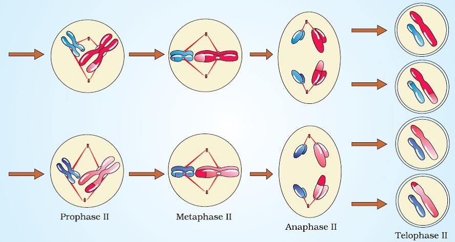
Prophase II
- Unlike prophase I, the prophase II is very brief.
- The nuclear membrane breaks down and nucleolus disappears.
- The chromatids undergo condensation. They become thick and short.
- Centrioles begin to move towards opposite poles.
- The spindle fibres develop.
Metaphase II
- Centrioles establish the poles of the spindle.
- The spindle will be at right angles to the plane of the spindle of metaphase I
- The kinetochores of the sister chromatids get attached to the opposite poles of the spindle through the chromosomal fibres and chromosomes are brought to the equatorial plate by congression.
- The chromosomes move and occupy the equator of th spindle. Unlike in metaphase I, they form a single metaphase plate with centromeres on equator and arms facing the poles.
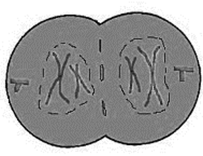
Anaphse II
- The centromere of each chromosome divides
- The sister chromatids become daughter chromosomes
- The chromosomal fibers shorten and carry the daughter chromosomes towards the opposite poles. The arms of the chromosome follow the centromere.
Telophase II
- The chromosomes undergo de-condensation. They become thin and long.
- The chromosomes form a network of chromatin.
- Spindle fibres disappear.
- Nuclear envelop is formed around the chromatin mass and nucleolus reappears.
Stages of Meiosis II
The telophase II is followed by cytokinesis. After karyokinesis two daughter cells undergo simultaneous cytokinesis by cleavage. In plant cells wall material is deposited between protoplast and a tetrad of haploid cells are formed. Thus, at the end of meiosis four haploid daughter cells with genetic variations are produced.
6. SIGNIFICANCE OF MEIOSIS
- Maintenance of constancy of chromosome number. Meiosis help in the maintenance of a constant number of chromosomes specific to a species. It reduces the diploid number of chromosomes in the gamete forming cell (germ cells) to haploid number in the gametes. Thus, when the gametes of sexually reproducing organisms fuse a diploid zygote is formed.
- Meiosis introduces genetic variations. During meiosis crossing over occurs resulting in new combination of genes among the daughter chromosomes. Even the paternal and maternal chromosomes assort independently. This leads to different combination of paternal and maternal chromosomes among the daughter cells. The genetic variations help in the evolution of species.
- Meiosis is essential for the formation of gametes in the sexually reproducing organisms.
- Meiosis may introduce chromosomal mutations sometimes harmful.
6. DIFFERENCES BETWEEN MITOSIS AND MEIOSIS
|
Characters |
Mitosis |
Meiosis |
|
I. GENERAL |
||
| 1. Site of occurrence
2. Period of occurrence 3. Nature of cells 4. Number of divisions 5. Number of daughter cells 6. Nature of daughter cells |
Somatic cells and during the multiplicative phase of gametogenesis in germ cells.
Throughout life. Haploid or diploid Parental cell divides once. Two Genetically like parental cell. Amount of DNA and chromosome number is same as in parental cell |
Reproductive or Germ
cells of gonads. During sexual reproduction Always diploid. Parent cell divides twice. Four Genetically different from parental cell. Amount of DNA and chromosome number is half to that of parent cell. |
| II. PROPHASE | ||
| 7. Duration
8. Subphases
9. Bouquet stage 10. Synapsis
11. Chiasma formation and crossing over 12. Disappearance of nucleolus and nuclear membrane 13. Nature of coiling |
Shorter (of a few hours) and simple.
Formed of 3 subphases: early-prophase, mid-prophase, and late-prophase. Absent Absent
Absent
Comparatively in earlier part. Plectonemic |
Prophase-I is very long (may be in days or months or years) and complex
Prophase-I is formed of 5 sub-phases- Leptotene, Zygotene, Pachytene, Diplotene and Diakinesis Present in Leptotene stage. Pairing of homologous chromosomes in Zygotene stage. Occurs during Pachytene stage of Prophase-I Comparatively in later part of Prophase-I. Paranemic. |
|
III. METAPHASE |
||
| 14. Metaphase plates
15. Position of centromeres
16. Number of chromosomal fibres |
Only one equatorial plate.
Lie at the equator. Arms are generally directed towards the poles. Two Chromosomal fibres join at centromere. |
Two plates in Metaphase-I but one plate in Metaphase-II
Lie equidistant from equator and towards poles in Metaphase-I while lie at the equator in Metaphase-II. Single in Metaphase-I while two in Metaphase-II |
|
IV. ANAPHASE |
||
| 17. Nature of separating chromosomes
18. Splitting of centromeres and development of inter-zonal fibres |
Daughter chromosomes (Chromatids with independent centromeres) separate.
Occurs in Anaphase. |
Homologous chromosomes separate in Anaphase-I while chromatids separate in Anaphase-II
No splitting of centromeres. Inter-zonal fibres are development in Metaphase |
|
V. TELOPHASE |
||
| 19. Occurrence | Always occurs | Telophase-I may be absent, but Telophase-II is always present. |
|
VI. CYTOKINESIS |
||
| 20. Occurrence
21. Nature of daughter cells
22. Fate of daughter cells |
Always occurs.
2N amount of DNA than 4N amount of DNA in parental cell. Divide again after interphase. |
Cytokinesis-I may be absent, but Cytokinesis-II is always present.
1N amount of DNA than 4N amount of DNA in parental cell. Do not divide and act as gametes. |
| VII. SIGNIFICANCE | ||
| 23. Functions
24. Variations
25. In evolution |
Helps in growth, healing, repair, and multiplication of somatic cells. Occurs in both asexually and sexually reproducing organisms.
Variations are not produced as it keeps quality and quantity of genes same. No role in evolution. |
Produces gametes which help in sexual reproduction. Occurs in only sexually reproducing organisms.
Produces variations due to crossing-over and chance arrangement of bivalents at Metaphase-I. It plays an important role in speciation and evolution. |
Formulae Chart
| 1. | Number of mitotic divisions for the formation of n number of cells. Example: For getting 100 cells 99 mitotic divisions are required. | n – 1 |
| 2. | Number of generations (n) of mitosis for producing ‘x’ cells. | x = 2n |
| 3. | Number of meiosis for the formation of ‘n’ seeds/grains/fruits. |