


1. INTRODUCTION
- A Three dimensional map of brain’s functioning can be obtained with FMRI (Functional Magnetic Resonance imaging) and a computer
- Among the vertebrates the nervous system reached the peak of evolution in man, especially with the development of the neopallium and the corpus callosum
- Human brain remains an Ultra-computer, the working mechanism of whose processor has not yet been deciphered or may be it can never be deciphered at all
- The study of Nervous system and its disorders is called Neurology
- Research work is going on to find out why people undergo dementia, involving memory loss as seen in Alzheimer’s .in the aging process.
2. CONTROL AND COORDINATION
- In human beings all physiological functions are coordinated by both the neural and endocrine systems.
- The neural system serves the following basic functions
(i) Receiving sensory inputs from internal and external environment by nerves to the brain.
(ii) Processing the input information by a central processor, the brain
(iii) Responding to stimuli transmitting motor commands from the brain to determine the response of the body parts or cells. - The endocrine system provides chemical integration through
- Coordination is the process in which two or more organs interact and complement the functions of one another.
- Coordination among the organs/organ systems in our body is a must to maintain homeostasis.
eg :
(1) During physical exercise muscles demand more energy which necessitates increased supply of oxygen by increased rate of respiration, heart beat and increased flow of blood in blood vessels.
(2) When physical exercise is stopped the activities of neural, lungs, heart and kidney gradually return to their normal conditions. - In our body neural system and the endocrine system jointly coordinate and function in a synchronised fashion.
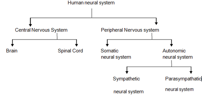
3. HUMAN NEURAL SYSTEM
- The human neural system has two sub systems, the central nervous system (CNS) and the peripheral nervous system (PNS).
- The central nervous system consists of a complex brain that is continuous with the dorsal tubular spinal cord.
- Peripheral nervous system comprises the nerve fibres.
- Nervous fibres are of two types
- afferent (sensory) fibres
- efferent (motor) firbres
- Sensory (afferent) nerve fibres : transmit impulses from tissues/organs to the CNS
- Motor (efferent) nerve fibres : transmit regulatory impulses from the CNS to the concerned peripheral tissues/organs.
- PNS is divided into two divisions
a) somatic neural system
b) autonomic neural system - Somatic neural system relays impulses from CNS to skeletal / Striated/voluntary muscles
- Autonomic neural system transmits impulses from the CNS to involuntary organs and smooth muscles.
- Autonomic neural system is further classified into
a) sympathetic neural system
b) parasympathetic neural system
4. CENTRAL NERVOUS SYSTEM
- Central nervous system includes brain and spinal cord. It develops from neurectoderm.
Brain
- Brain is the central information processing organ of our body, and acts as the ‘command and control system’.
- The brain is situated inside the cranium and it is covered by three connective tissue membranes called cranial meninges.
- The three layers of meninges are the outermost dura mater, middle archnoid membrane and inner pia mater in contact with the brain tissue
- Arachnoid mater is seperated from the dura mater by the subdural space and from pia mater by subarachnoid space containing cerebrospinal fluid.
- The human brain can be divided into forebrain, midbrain and The hindbrain continues into the spinal cord.
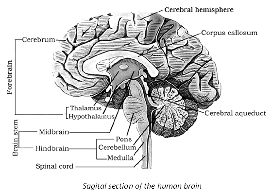
Fore Brain
- The forebrain consists of 3 parts
- Olfactory bulbs
- Cerebrum
- Diencephalon
Olfactory Bulb :- Olfactory bulbs receive impulses pertaining to smell from the olfactory epithelium.
Cerebrum
- Cerebrum forms major part of brain.
- The cerebrum is divided into two hemispheres by prominent longitudinal fissure. The two hemi-spheres are connected by a bundle of trans-verse fibres called corpus callosum. or colossal commissure The left half of the brain controls the right side of the body and vice versa.
- Each cerebral hemisphere is divided by other grooves into four lobes namely, frontal, tem-poral, parietal and
- Cerebral cortex is the outer layer of the It is made of grey matter. Neuronal cell bodies are concentrated in cerebral cortex.
- The surface of each cerebral hemisphere shows many convolutions called gyri (singular; gyrus) the deepest and shallower grooves between the folds are called fissures and sulci respectively.
- The sulci and gyri increase the surface area of the cortex.
- In cerebral hemispheres three types of functional areas are present.
- Sensory areas receive and interpret sensory impulses from receptors.
- Motor areas which control voluntary muscular movements.
- Association areas which are neither clearly sensory nor motor in function and they deal with more complex ‘integrative functions’ such
Diencephalon (Thalamencephalon)
- The main parts of the diencephalon are the epithalamus. thalamus and hypothalamus
- Epithalamus : It is the roof of the dincephalon. It is a non-nervous part which is fused with the pia mater to form the anterior choroid plexus.
- Just behind the anterior choroid plexus, the epithelium of the epithalamus forms a pineal stalk, which ends in a rounded structure called pineal body.
- Thalamus : It lies superior to the mid brain. It is the major coordinating centre for sensory and motor signalling.
- Hypothalamus (the thermostat of the body) :
It lies at the base of the thalamus. - The hypothalamus forms a funnel shaped down-ward extension called infundibulum connecting the hypothalamus with the pituitary gland.
- Hypothalamus contains neural centres for hunger, thirst, satiety and groups of
neurosecretory cells, which secrete hormones called hypothalamic hormones. - Hypothalamus controls and integrates the activities of the autonomous nervous system.
- Limbic system : The inner parts of the cerebral hemispheres and a group of associated deep structures like the hippocampus, the amygdala, etc.
- The limbic system along with hypothalamus is involved in the regulation of sexual behaviour and expression of emotional reactions.
Mid Brain (Meseccephalon)
- Mid brain is located between thalamus/hypo thalamus of the fore brain and pons of the hind brain.
- A canal called the cerebral aqueduct passes through the mid brain.
- It contains four rounded lobes, the corpora quadrigemina. Its chief structures are superior colliculi and inferior colliculi.
- The anterior, larger superior pair of colliculi are concerned with vision.
- The posterior, smaller inferior pair of colliculi are concern with auditory functions.
- The ventral portion of the midbrain consists of a pair of longitudinal bands of nervous tissue called cerebral peduncles or Crura cerebri.
Hind Brain or Rhombencephalon
- The hind brain comprises pons, cerebellum and medulla (Medulla oblongata).
Cerebellum
- The cerebellum is the second-largest part of the brain. It is wedged between cerebral hemi-spheres and brain stem. The cerebellum is divided into two hemispheres and central
- Each cerebellar hemisphere consists of three lobes namely anterior, posterior and floccular lobes.
- The surface of each hemisphere is made up of gray matter surrounding a large mass of white matter .
- The cerebellum is solid. It has a braching tree-like core of white matter called arborvitae surrounded by a sheath of grey matter.
- The cerebellum is vital to the control of rapid muscular activities, such as running, typing and even talking.
Pons varolii
- Pons appears as a rounded bulge on the underside of the brain stem.
- It separates the midbrain from the medulla oblongata.
- It consists of nerve fibres which form a bridge between the two cerebellar hemispheres
- It is a relay station between the cerebellum, spinal cord and the rest of the brain.
- Pneumotaxic area and apneustic area are involved in the control of the respiratory muscles
- Pons serves as a neuronal link between the cerebral cortex and the cerebellum.
Medulla Oblongata
Medulla oblongata passes out of the foramen magnum and joins the spinal cord
- It has a very thin , vascular folded structure called posterior choroid plexus
- The medulla oblongata is the lower portion of the brainstem. It is inferior to the pons. It controls autonomic functions like respiration, cardiovascular reflexes and gastric secretions, swallowing vomiting, sneezing, hiccuping etc.
Brain Stem
- The medulla oblongata, the pons, and mid-brain lie in a portion of the brain known as brain stem connecting the fore brain and the spinal cord.
- These structures include many tracts of nerve fibres and masses of grey matter called nuclei.
Ventricles of The Human Brain
- Human brain consists of four ventricles.
- The first and second ventricles ( lateral ventricles or paracoels) are present in the right and left cerebral hemisheres respectively.
- The two paracoels are connected to the median diocoel individually by the two foramina of Monro ( Interventricular foramina)
- The third ventricle ( diocoel) occurs in the
- The fourth ventricle ( myelocoel) is present in the medulla.
- The myelocoel and the diocoel are connected by a narrow canal called iter or aqueduct of sylvius/ cereral aqueduct.
- The metacoel is continuous with the central canal of the spinal cord
- The ventricles of the brain, and the subrachnoid space are filled with Cerebro-spinal fluid (CSF) .
- Cerebro-spinal fluid (CSF) . is an alkaline, colourless fluid which is filtered from the choroid plexuses into the ventricles of the brain
- CSF serves as shock absorbing medium CSF is recycled (flushed) 4 times per day in order to clear out metabolites and toxins
IMPORTANT FUNCTIONS OF BRAIN
| FOREBRAIN | |
| Olfactory region Cerebrum |
Smell Thinking, Intelligence, Memory, Ability to learn Reasoning, Conscious, Control, Speech micturition (passing of urine) Defecation, voluntary forced breathing, voluntary muscular coordination etc. |
| Diencephalon (hypothalamus) |
Integrates the activities of the autonomic nervous system, has control centres for hunger. Thirst, Sweating, appetite hunger, satiety etc. and regulate body temperature so thermostat of the body. |
| MIDBRAIN | Reflex centre of visual and auditory sensation |
| HINDBRAIN | |
| Cerebellum | Involuntary muscular coordination. Maintain posture, Orientation and equilibrium of the body. |
| Medulla Oblongata | Regulate heart rate, involuntary breathing respiratory centre, Blood pressure (Vasoconstriction and vasodilation). Gut peristalsis, food swallowing, vomiting, Gland secretion. |
Spinal Cord
- Spinal cord is located in the vertebral canal (neural canal) of the vertebral column. It is an elongated cylindrical structure which lies in the neural canal of the vertebral column and is continued with the medulla oblongata through foramen magnum of the skull.
- In the adult, it extends from the medulla oblongata to the superior border of the second lumbar vertebra.
- In the neck region, a thickening in the spinal cord is called the superior cervical enlargement.
- Inferior enlargement in the Spinal cord is called the lumbar enlargement
- Just inferior to the lumbar enlargement, The spinal cord tapers to a conical portion known as the conus medullaris.
- Conus medullaris ends at the level of the intervertebral disc between the first and second lumbar vertebrae in an adult.
- The extension of the conus medullaris as the non-nervous fibrous tissue to the coccyx is called ‘filum terminale’.
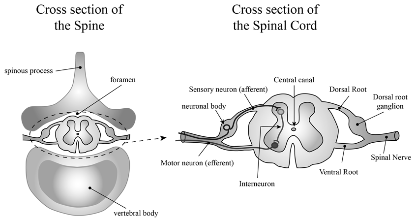
Internal anatomy of spinal cord
- Unlike the brain, grey matter of the spinal cord is located centrally, surrounded by outer white matter.
- In a cross section, spinal cord consists of a H or a butterfly shaped central core of grey matter surrounding a central canal and an outer layer, the white matter. The spinal cord is divided into right and left halves by two grooves namely
a) posterior, median dorsal sulcus
b) Anterior ventral median fissure - Narrow longitudinal cavity of Spinal cord is called central canal.
- central canal is lined by the ependymal epithelium, continues with 4th ventricle of medulla oblongata.
- The grey matter is subdivided into regions called ‘anterior and posterior horns’.
White matter: White matter consists of bundles of myelinated axons and it is organized into the regions called anterior (ventral) funiculus, posterior (dorsal) funiculus and lateral funiculi. - The spinal cord acts as a coordinating centre for simple spinal reflexes. It also acts as the
- ‘middle man’ between the receptors and the effectors
5. PERIPHERAL NERVOUS SYSTEM (PNS)
- It consists of nerves connected to or arising from the central nervous system.
- It has cranial and spinal nerves.
Cranial Nerves
- In man 12 pairs of cranial nerves are present.
- In all anamniotes the total no of cranial nerves are 10 pairs.
- In all amniotes the total no of cranial nerves are 12 pairs except snakes.
Note : Refer to chart
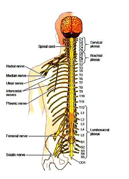
Spinal Nerves
- Spinal nerves arise from the grey matter of spinal cord.
- There are 31 pairs of spinal nerves in man.
- Spinal nerves are mixed in nature because each spinal nerve is formed of 2 roots
a) sensory (afferent) root
b) motor (efferent) root. - Each spinal nerve is divided into three rami
a) Ramus dorsalis
b) Ramus ventralis
c) Ramus communicans – it joins sympathetic ganglion of autonomic nervous system. - The spinal nerves in man are divided into five groups
- Spinal nerves in man – 31 pairs
1) Cervical nerves [C] – 8 pairs – neck region.
2) Thoracic nerves [T] – 12 pairs – thoracic region.
3) Lumbar nerves [L] – 5 pairs – upper part of abdomen.
4) Sacral nerves [S] – 5 pairs – lower part of abdomen.
5) Coccygeal nerves [CO] – 1 pair - Spinal nerve formula can be written as C8,T12,L5,S5,CO1.
- The first pair of cervical spinal nerves emerge between the atlas and occipital bone of the
- All other spinal nerves emerge from the vertebral column through the intervertebral foramina
- The lumbar, the sacral and the caudal nerves extend back along with the filum terminale forming a thick bundle of nerves called cauda equina
- Certain spinal nerves are joined to form networks called plexuses
- 1. Cervical plexus( 1st to 4thcervical nerves)
2) Brachial plexus ( 5th to 8th cervical and Ist thoracic nerves)
3) lumbar plexus (1st lumbar and a branch h of the 4 lumbar nerve)
4) Sacral plexus ( The 1st three Sacral a branch of the 4th lumbar and the 5th lumbar nerves)
5) Coccygeal plexus (4th and 5th sacral and coccygeal nerve).
CRANIAL NERVES OF Man AT A GLANCE
| S.No | Name | Nature | Origin | Distribution | Function |
| I | Olfactory Nerves | Sensory | Olfactory lobe | Sensory epithelium of olfactory sacs | Receive stimuli from the sensory epithelium of olfactory sac and carry them to olfactory lobes |
| II | Optic nerves | Sensory | In retina of eye | Lateral geniculate nuclei of thalamus | Stimulus of light is carried to optic occipital lobe of cerebral cortex |
| III | Occulomotor nerves | Motor | Crura cerebri (mid brain) | Eye ball muscles like superior rectus, medial rectus, inferior rectus and inferior oblique, except superior oblique muscle and external rectus | Movement of eye lids and eye ball |
| IV | Trochlear nerves | Motor | From in between the optic lobes and cerebellum | Superior oblique muscle of eye ball | Movement of eye ball |
| V | Trigeminal nerves | Mixed | From the gassarion ganglia situated on the lateral side of pons | – | – |
| (i) Ophithalimic Nerve | Sensory | “ | Skin of lips, upper eye lid, lacrimal, gland | ||
| (ii)Maxillary | Sensory | “ | Upper lip, skin of nose, lower eye lid. Upper teeth | Carry the stimuli from these organs to brain | |
| (III) Mandibular Nerve | Mixed | “ | Lower lip and skin of Jaw | Carry the stimuli from these organs to brain | |
| VI | Abducens nerves | Motor | Pons | Eye muscles external rectus | Movement of eye ball |
| VII | Facial nerves | Mixed | Pons | – | – |
| (i) Palatinus | Sensory | – | In the roof of mouth cavity | Carry the impulses from roof of mouth cavity | |
| (ii) Hyomandibular | Motor | – | Muscles of lower jaw, muscles of neck and pinna (external ear) | Carrythe impulses from brain muscles of lower jaws, neck and pinna | |
| (iii) Chordatympani | Mixed | – | In salivary glands and taste buds | Receives the stimuli from the taste buds and carry the stimulus to salivary gland | |
| VIII | Auditory nerves | Sensory | Medulla, pons | – | – |
| (i) Vestibular nerve | Sensory | – | Semicircular canals, saccule, utricle. | Receives impulses from the internal ear and carry to brain for equilibrium | |
| (ii) Cochlear nerve | Sensory | – | Cochlea | Impulses associate with hearing | |
| IX | Glossopharyngeal nerve | Mixed | In medulla | Taste buds present in tongue and muscles of oesophagus | Secretion of saliva, taste muscle sense (proprioception) |
| X | Vagus nerve | Mixed | Arising from medulla, 9th and 10th cranial nerves unites to form vagus nerve but become separate and divide into branches | – | – |
| (i) Superior laryngeal nerve | Motor | – | Glottis, trachea, lung muscle | (1) Smooth muscles contraction and relaxation (2) Secretion of digestive juice (3) Muscle sense (proprioception) (4) Sensation of visceral Organs |
|
| (ii) Recurrent Laryngeal nerve | Motor | – | Glottis, trachea, lung muscle | ||
| (iii) Cardiac nerve | Motor | Heart Muscles | From brain to heart muscles | ||
| (iv)Pneumogastric | Motor | – | In the abdominal cavity, in stomach and lungs | Carry impulse from these organs to brain and from brain to muscles of these organs | |
| (v) Depressor nerve | Motor | – | Diaphragm | Carry the impulse to diaphragm | |
| XI | Spinal accessory | Motor | Medulla | Muscles of neck and shoulders, voluntary muscles of pharynx, larynx, and soft palate | Swallowing movements, movement of head |
| XII | Hypoglossal nerve | Motor | Medulla | Muscles of tongue and neck | Movement of tongue during speech, and swallowing, proprioception (Muscle sense) |
Functionally, the PNS is divided into two divisions called Somatic and Autonomic neural systems.
Somatic neural system (SNS)
- SNS includes both sensory and motor neurons.
- From somatic receptors the sensory neurons conduct sensory impulses to CNS.
- These sensations are consciously perceived.
- The motor neurons of SNS have single myelinated axons and they innervate the skeletal muscles and produce voluntary movements.
- The result is always excitation
6. AUTONOMIC NERVOUS SYSTEM
- ANS controls and coordinates the activities of visceral organs by controlling visceral sensation and visceral movements.
- Autonomic nervous system usually operates without conscious control.
- Autonomic nervous system is entirely motor.
- All autonomic axons are efferent fibres.
- The centres in brain like cerebral cortex, hypothalamus and medulla oblongata regulates the autonomic nervous system.
- ANS consists of two divisions
a) sympathetic division
b) para sympathetic division
Sympathetic Division
- The preganglionic neurons arise from the thoracic and lumbar regions of the spinal cord, hence it is called as thoracolumbar
- It consists of two sympathetic chains one on either side of the dorsal aorta and beneath the vertebral column on either side of dorsal aorta extending from base of the skull to the pelvis of the body.
- The axons of sympathetic preganglionic neurons are called thoracolumbar outflow.
- In sympathetic division preganglionic nerves are small where as post ganglionic nerves are long.
- Each sympathetic chains bears a series of trunk ganglia/chain ganglia Besides trunk ganglia,
- three collateral ganglia lie outside sympathetic chain close to large abdominal arteries below diaphragm
- Collateral ganglia are coeliac, superior mesentric and inferior mesenteric ganglia.
- Preganglionic nerve secretes acetyl choline and postganglionic nerve secretes sympathin (nor-epinephrine)
Parasyampathetic Division
- It is called as cranio-sacral division.
- The parasympathetic neural system is said to exhibit ‘Cranio-sacral outflow’
- Cranial nerve fibres of III,VII,IX and X along with 2,3,4 sacral nerve fibres form parasympathetic nervous system.
- Parasympathetic ganglia lie in head, neck and sacral region.
- Preganglionic nerve is long whereas postganglionic nerve is small.
- Parasympathetic ganglia are located close to or within the wall of visceral organs. Hence they are called terminal ganglia.
- These nerve fibres secrete acetyl choline only.
- The sympathetic and parasympathetic nervous systems are antagonistic in their action.
- Sympathetic nerve fibres are adrenergic while parasympathetic nerve fibres are cholinergic in their mode of action.
7. COMPARISON OF SOMATIC AND AUTONOMIC
| Nervous Systems | Somatic Nervous System | Autonomic Nervous System |
| Sensory input | From somatic senses and special senses | Mainly from interoceptors |
| Control of motor output | Voluntary | Involuntary |
| Motor neuron pathway | One-neuron pathway | Two neuron pathway |
| Neurotran- smitter | Ach | Ach and NE |
| Effectors | Skeletal muscles | Smooth muscles, cardiac muscles and glands |
8. FUNCTIONS OF SYMPATHETIC AND PARASYMPATHETIC NERVOUS SYSTEM
|
S.No |
Name |
Sympathetic |
Parasym pathetic |
|
1.
2. 3. 4. 5. 6. 7. 8. 9. 10
11. 12. 13. 14. 15 |
Secretion
Blood pressure Blood Vessel to heart Pupil Lacrimal gland Heart beat Adrenal Secretion Breathing and BMR Sphincters Urinary Bladder
Salivary secretion Digestive secretions Gut Peristalsis Bronchi Gastrointestinal motility |
Acetyl choline and Sympathin
Increases Dilates Dilates Stimulates Increases Stimulates Increases Contracts inhibits emptying bladder Decreases Decreases Decreases Dilate Decreases |
Acetyl Choline only Decreases Constricts Constricts Inhibits Decreases Inhibit Decreases Relaxes Promotes emptying bladder Increases Increases Increases Constrict Increases |
9. GENERATION AND CONDUCTION OF NERVE IMPULSE
- Neuron is the structural and functional unit of Neural System
- Neuron membrane is in a polarised state hence neurons are excitable cells.
- Nerve cell exhibit special property called electrical excitability
- The signal that travels along the length of a nerve fiber and ends in the release of neurotransmitters is called a nerve impulse
- On the neural membrane different types of ion channels are present making it polarised.
- The axolemma of a neuron has three different types of ion channels
- Leakage channels : They are Na+ and K+ leakage channels
- The axolemma has many K+ leakage channels than Na+ leakage channels.
- Only one gate is present in voltage – gated K+ channel
- Ligand-gated channels : They are present on post-synaptic membrane and they open or close in response to a chemical stimulus.
- Voltage-gated channels : They are responsible for the generation of membrane potentials
- There are two types of voltage -gated channels one for Na+ and another for K+.
- Sodium has activation and inactivation voltage – gated channels.
- K+ has only one activation voltage gated channel.
Resting Membrane Potential
- When a nerve fibre is at rest, its outer surface is positively charged and its inner surface is negatively charged due to a small buildup of positive ions in the ECF and equal buildup of negative ions in the axoplasm
- Such a separation of positive and negative electrical charges is a form of potential energy.
- At resting face the resting membrane potential changes from – 40 to -90 mV
- A typical value is – 70 mV.
- If the inner side becomes less negative, it (axolemma) is said to be depolarized. {It means more than – 70 mV like – 55mV}
- When the side is more negative then it is said to be hyperpolarised. {from -70mV to -90mV}
- When the voltage – gated Na+ channel is in resting condition, the activation gate is closed and the inactivation gate is open
- When the channel is activated, both the gates are open
- The membrane potential of neuron that is not transmitting signals is called the resting potenial
- Resting potential depends on the ionic gradients and the differential permeability of the axolemma
Sodium -potassium pump
- The Na+ and K+ gradients are maintained by the sodium -potassium pump ( Na+/K+ AT- Pase)
- Sodium potassium pump transports three Na+ into ECF and two K+ into the cell.
Important Note
- As these pumps remove more positive charges from the axoplasm than they bring into it, they
- contribute to the negativity of the resting membrane potential i.e., – 70mv.
- At the resting potential, the voltage – gated Na+channels are in resting state. Voltage – gated K+ channels are closed.
Depolarization (Rising phase)
- When a nerve fibre is stimulated, the plasma membrane becomes more permeable to Na+ ions than to K+ during these activation gate for Na+ is opened. K+ gate is closed.
- Due to the influx of Na+axolemma is positively charged inside and negatively charged outside. This reversal of electrical charge is called “depolarization”.
- The electrical potential difference between two areas is called “action potential”.
- Action potential occurs when depolarization reaches threshold level – 55mV.
- At the peak of depolarization the potential is +45mV. (spike potential)
- Action potential is an all – or – none phenomenon all – or – none principle :-
- The action potential occurs in response to a threshold stimulus or supra threshold stimulus but does not occur at subthreshold stimuli. It means the nerve impulse is either conducted totally or not conducted at all and this is called ‘all – or – none principle’.
Repolarization (Falling phase)
- When the membrane potential rises to its peak the voltage gated Na+ channels are inactivated and voltage gated K+ channels open
- Efflux of K+ repolarizes the axolemma.
Hyper Polarization (under shoot) During hyperpolarizing phase, the membrane potential becomes – 90mV.
Voltage gated K+ channels are open, activation and inactivation voltage gated Na+ channels remain close
| State | Sodium voltage gated channels |
Potassium voltage gated channels |
|
| Activated | Inactivated | Activated | |
| Resting Depolarization | Closed Open |
Open Closed |
Closed Closed |
| Repolarization | Open | Close | Open |
| Hyper polarization | Close | Close | Open |
The Refractory Periods
- The period of time after an action potential begins during which the neuron cannot generate another action potential in response to a normal threshold stimulus is called the ‘refractory period’
- Refractory periods are of two types
- The few milliseconds period after initiation of an action potential, during which it is impossible to trigger a second action potential is known as the absolute refractory period
- During absolute refractory period voltage-gated Na+ channels are either activated (depolarizing phase) or inactivated (repolarizing phase).
- The time period during which a second action potential can be initiated by a larger than normal stimulus is termed relative refractory period
- During relative refractory period voltage -gated K+ open and Na+ channels are in resting state ( hyperpolarizing phase)
Propagation of Action Potentials
- Action potential is initiated at axon hillock.
- Action potential cannot be generated in the zone behind the travelling zone of depolarization as it is in refractory period
- Conduction speed is directly proportional to the diameter of axon.
- In a myelinated axon, volatage – gated Na+ and K+ channels are concentrated at the nodes of
- Type of conduction in myelinated axons is saltatory conduction.
- Saltatory conduction is faster than the continuous conduction because the action potential reaps from node to node
Synaptic Transmission
- Transmission of nerve impulses are carried through synapse.
- A synapse is a junction between two neurons or between a neuron and an effector (muscle or gland).
- A synapse is formed by
- presynaptic membrane
- postsynaptic membrane.
- Synapse may or may not be separated by a gap called Synaptic Cleft.
- There are mainly two types of synapses:
- electrical
- chemical, depending upon the nature of transfer of information across the synapse.
- Electrical Synapses: These are specialized for rapid signal transmission. It is similar to impulse conduction along a single axon.
- At electrical synapses, the membranes of pre-and post-synaptic neurons are in close proximity, with gap junctions
- Electrical currents can flow directly from one neuron to other across these synapses which are faster than chemical synapses.
- Electrical synapses are rare in our body system.
- Chemical synapses: The majority of synapses are chemical synapses. Presynaptic and post synaptic neurons are separated by a fluid filled space, the synaptic cleft (structural gap and a functional bridge)
- It consists of a bulbous expansion of a nerve terminal, called synaptic knob, lying in close proximity to the membrane of a dendrite.
- The cytoplasm of the synaptic knob contains numerous tiny, round sacs, called synaptic vesicles, which contain neurotransmitter substance responsible for the transmission of nerve impulse across the synapse.
- When a wave of depolarisation reaches the presynaptic membrane, voltage-gated calcium channels, concentrated at the synapse open.
- Ca2+ ions, enters into knobs and stimulate synaptic vesicles in the terminal to move to the terminal membrane, fuse with it and there by releasing neurotransmitter chemicals from vesicles into the cleft, by exocytosis.
- The post synaptic membrane has ligand gated ion channels
- These neurotransmitters rapidly pass to the other side of the gap. They then combine with specific receptor molecules on the membrane of the target cell, which is called the postsynaptic membrane. By doing so, they cause small and short-lived electrical signals in the dendrite of the second neuron.
The new potential developed may be either excitatory or inhibitory. - Excitatory neurotransmitters depolarize the postsynaptic membrane and produce excitatory postsynaptic potentials(EPSPs).
- Inhibitory neurotransmitters hyperpolarize the postsynaptic membrane and produce inhibitory postsynaptic potentials (IPSPs).
- Action potentials are all-or-none, but postsynaptic potentials are graded
- Graded potentials do not have refractory period and undergo summation
- The integration of these inputs from many synapses is known as
- Summation of postsynaptic potentials occurs at axon hillock.
- The summation of inputs from several presynaptic boutons is called spatial summation
- The summation of successive inputs from a single presynaptic bouton is called temporal
summation
- An action potential is generated in the initial segment of the axon of the postsynaptic neuron if the sum of all the EPSPs exceeds the sum of IPSPs and the threshold is reached. Most common neurotransmitter both in invertebrates and in vertebrates is Acetylcholine.
- Acetylcholine is either excitatory or inhibitory, depending on the type of receptor
- Enzyme that degrades acetylcholine is acetylcholinesterase
- Acetylcholinesterase resides in the synaptic cleft Some biogenic amines involved in synaptic transmission are epinephrine, norepinephrine. dopamine and serotonin. They are excitatory or inhibitory Amino acids that act as only inhibitory neurotransmitters are Gamma aminobutyric acid (GABA) and glycine.
- Wave of depolarisation reaches the presynaptic membrane.
- Voltage – gated calcium channels open, Ca++ ions diffuses into the axon terminal from the surrounding fluid
- Ca++stimulates fusion of synaptic vesicle with pre-synaptic membrane, and release of neurotransmitter by exocytosis into synaptic cleft.
- Neurotransmitter binds with specific receptor molecules of post – synaptic membrane
- This binding opens specific ion channels allowing the entry of specific ions which can generated a new potential in the post – synaptic neuron
- The new potential developed may be either excitatory or inhibitory depending upon neurotransmitter.
10. REFLEX ACTION AND REFLEX ARC
- A reflex action is a quick autonomic response to a stimulus. The response does not involve
- any conscious effort. Therefore, reflexes are described as involuntary actions.
- A spinal reflex is a reflex in which integration takes place in the spinal cord.
- A cranial reflex is reflex in which integration takes place in the brain stem rather than spinal cord.
- The tracking movements of your eyes as you read this sentence.
- The pathway followed by nerve impulses that produces a reflex is called a reflex arc. The response is called a reflex.
- The simplest reflex arc involves some specific
1) receptor(s), 2) afferent sensory neuron(s), 3) Internuncial neuron
4)efferent neuron 5) effector - The terminal dendrites of the afferent sensory neuron lie in a dorsal root ganglion of the spinal cord. The dendrities of the efferent motor neuron lie in a ventral root.
- Stimulation of the receptors initiates a nerve impulse along the afferent neuron connected to them.
- The nerve impulse flows along the afferent, connector and efferent neurons to reach a muscle
- or gland, called an effector for that reflex.
- This produces either a movement of the muscle or the secretion of the gland as the effect of the
- Coughing is a reflex action. The stimulus is a foreign matter in the respiratory tract .
 Sensory Reception and Processing
- Sensation is the conscious or subconscious awareness of changes in the external or internal environment. In the human body the different types of receptors detect all types of changes in the environment and send appropriate signals to the CNS, where all the inputs are processed and analysed. Then the different controlling centres of the brain send ‘motor impulses’ to the ‘effectors’ through motor nerves.
11. IMPORTANT RECEPTORS OF HUMAN BODY
- Exteroceptors: They are located at or near the surface of the body, and are sensitive to external stimuli like hearing, light, touch, taste and pain etc.
- Interoceptors: They are located in the visceral organs and blood vessels and sensitive to internal stimuli.
- Proprioceptors: They are also a kind of interoceptors and they provide information about body position and movement and are located in muscles, tendons, joints and the internal ear.
- Thermoreceptors : They respond to heat (caloreceptors) and cold (frigidoreceptors)
12. EYE
- The eye is important and delicate structure. The orbits called bony sockets in the skull will protect the eyes from physical damage.
Parts of An Eye
1) accessory structures
2) Eye ball
Accessory structures :- Eye lids, eye lashes, eye brows, lacrimal apparatus and extrinsic eye muscles are the accessory structures of eye. The eye lids, the eye lashes and the eye brows are useful for the protection of the eye. The lacrimal apparatus is a group of structures that produce and drain lacrimal fluid or tears which contains salts, mucus and bactericidal enzyme called lysozyme.
Eye Muscles
|
Eye muscles |
Innervated by |
|
Superior rectus Inferior rectus Lateral rectus Medial rectus Superior oblique Inferior oblique |
Oculomotor (III) nerve Oculomotor (III) nerve Abducens (VI) nerve Oculomotor (III) nerve Trochlear (IV) nerve Oculomotor (III) nerve |
- Adult human eye ball is nearly spherical in shape.
- Each eye ball is composed of three concentric
- layers They are
1. Fibrous tunic
2. Vascular tunic
3. Nervous tunic - Sclera and choroid are mesodermal. Rest of the eye is ectodermal.
- FIBROUS TUNIC :– It is the outer coat of eye ball
- It consisting of the anterior cornea (acts as a sort of ‘fixed lens’) and the posterior sclera.
Cornea
- The cornea is a non-vascular, transparent coat that covers the coloured iris.
- Cornea is covered by a thin layer called conjunctiva
Sclera
- The sclera ‘white’ of the eye, is a coat that covers the entire eye ball. The sclera gives shape to the eye ball, makes it more rigid, and protects its inner parts.
- At the junction of the sclera and cornea is a channel known as the sclera venous sinus or
- canal of Schlemm.
Vascular Tunic (Uvea)
- It is the middle layer of the eye ball.
- It has three portions :
1. CHOROID :- It is highly vascularised and - looks bluish in colour.
2. CILIARY BODY :- It is thick in the anterior part to form the ciliary body. - It is a pigmented and vascularised part that consists of the ciliary processes.
- The muscle associated with the ciliary body is a circular band of smooth muscle that holds and alters the shape of the lens for near or far vision.
3. IRIS :- It is the coloured portion of the eyeball. - It is suspended between the cornea and the lens and is attached at its outer margin to the ciliary processes.
- At the centre of iris is a an aperture called pupil Iris regulates the amount of light entering the eyeball through pupil.
Nervous Tunic (Retina)
- It is the third and inner coat of the eye.
- It consists of a pigmented epithelium (non-visual portion) and a neural portion .
- The neural portion of the retina has three layers of retinal neurons namely :
- 1) Photoreceptor layer (The layer closest to the choroid coat).
- 2) Bipolar cell layer
- 3) Ganglion cell layer
Photoreceptor Layer
- It consists of two types of photoreceptor cells called ‘rods’ and ‘cones’.
Rods :
- It contains a purplish-red protein called the rhodopsin or visual purple
- Rhodopsin contains a derivative of vitamin-A
- The rods are more sensitive towards light as compared to cones.
- Rods used for vision in dim light (scotopic vision), having no ability to detect colour.
Cones :
- It contain a visual pigment is called iodopsin, Iodopsin is made of a protein called Photopsin
- Cones are used for bright light vision with the ability to make coloured image of object.
- The cones are of three different types
1) Short wavelength-sensitive cones (blue)
2) Medium wavelength-sensitive cones (green)
3) Long wavelength-sensitive cones (Red)
Differences between Rods and Cones
| Rods | Cones |
|
Long and slender More numerous Uniformly distributed |
Shorter and thicker Less numerous Tightly packed at Fovea |
| One type | Three types (red, green, blue) |
| Contain rhodopsin Specialized for scotopic vision | Contain iodopsin Specialized for photopic |
| Sensitive to dim light | Sensitive to bright light |
| Cannot detect colours | Can detect colours |
| Less resolution | High resolution |
- Erythropsin, Chloropsin and Cyanopsin are respectively sensitive to red, green and blue.
- Colourblindness (daltonism) is due to the absence of one of the three types of cones.
Functions of Different Parts of Eye Fibrous Tunic Function
Sclera → Protects and supports eye ball
Cornea → Refracts light rays
Vascular Tunic
Choroid → Absorbs stray light
Ciliary body → Holds lens in place, accommodation
Iris → Regulates light entrance
Pupil → Admits light
Retina → Contains sensory receptors for light
Rod cells → Black-and-white vision
Cone cells → Colour vision
Fovea centralis → Acute vision
Others
Lens → Refracts and focuses light rays
Humors Transmit → light rays and support eye ball
Optic nerve → Transmits impulse to brain
Blind spot and Yellow spot :
- The centre of the posterior portion of the retina is called the macula lutea or yellow spot.
- A small depression present in the centre of yellow spot is called fovea centralis.
- Rods are absent in central fovea.
- Fovea is responsible for sharp, central vision, which is useful while walking, reading, driving etc.
- Macula lutea is placed above the blind spot (optic disc), from where the optic nerve emerges.
- At blind spot, neither rods nor cones are present and no image is formed. Present at the point of emerging of optic nerve.
DIFFERENCES BETWEEN BLIND SPOT AND YELLOW SPOT
| A region at the back of the eye where optic nerve exists Rods and cones are absent Image is not formed due to the absence of photoreceptor cells |
Yellowish area at the centre of the posterior portion of the retina Rods are absent in fovea centralis Vision is sharpest at fovea centralis |
Lens
- The lens is present within the cavity of the eye ball behind the pupil and the iris.
- Lens is a non-vascular and transparent
- The lens is composed of layers of proteins called
- The lens is held in position by encircling suspensory ligaments.
- Ciliary muscles control the shape of the lens helping focusing light on the retina.
Chambers of the eye’s interior
- The lens and its suspensory ligament divide the cavity of the eyeball into the anterior cavity and
- posterior cavities.
- The anterior cavity (aqueous chamber) is present between the cornea and lens.
- The anterior cavity consist of two chambers.
- The anterior chamber is present between cornea and iris
- The posterior chamber is present between iris and lens.
- The anterior chamber is filled with aqueous humor (secreted by ciliary processes),
- The aqueous humor helps in nourishing the lens and the cornea
- The posterior chamber is filled with a jelly like substance called vitreous body/vitreous humor.
- The vitreous body contributes to the intraocular pressure along with aqueous humor and helps to protect the shape of the eye ball.
Mechanism of Vision
- The light rays in visible wave length focused on the retina through the cornea and lens generate potentials (impulses) in rods and cones.
- Light induces dissociation of the retinol from opsin resulting in changes in the structure of the opsin. This causes membrane permeability changes. As a result, potential differences are generated in the photoreceptor cells. This produces a signal that generates action potentials in the ganglion cells through the bipolar neurons.
- These action potentials (impulses) are transmitted by the optic nerves to the visual cortex area of the brain, where the neural impulses are analysed and the image formed on the retina is recognised, based on earlier memory and experience.
13. EAR
- The human ear consists of three parts, the outer, middle and inner ear.
External Ear (Outer Ear)
- The outer ear consists of an immovable cartilaginous ear lobe or pinna, and an external au-ditory meatus (canal) leading inwards from it
- through cranial bones.
- The pinna is somewhat funnel-shaped. It collects and directs sound waves into the external auditory canal.
- The external auditory canal ends at a delicate membranous diaphragm, called eardrum or tympanic membrane.
- External auditory canal contains hair and specialized sebaceous glands called ceruminous glands.
- Ceruminous glands secrete cerumen or earwax.
- Ceruminous glands present in the skin of the pinna and the meatus.
- Hair and cerumen help to prevent dust and foreign particles from entering the ear
The Middle Ear
- The middle ear (tympanic cavity) is an air – filled chamber on the other side of the eardrum and inside the cranial bone.
- Middle ear cavity communicates with that of the pharynx through an air-filled tube, called eustachian tube.
- The Eustachian tube helps in equalizing the pressures of air on either sides of the ear drum.
- The middle ear separated from the internal ear by a thin bony partition that contains two small membrane – covered openings called the oval window (fenestra ovalis) and the round
window (fenestra rotunda). - The middle ear contains three ossicles called malleus (hammer bone), incus (anvil bone) and stapes (stirrup bone) which are attached to one another in a chain like fashion.
- Malleus is attached to the tympanic membrane and its head articulates with the incus
- The incus is the intermediate bone in the series of ‘ear ossicles’ and articulates with the head of the stapes.
- The Stapes is attached to the oval window in the thin bony partition between the middle and inner ear.
Internal Ear
- The fluid-filled inner ear is called labyrinth
1. Labyrinth consists of two parts
2. The bony labyrinth - The membranous labyrinth.
- The bony labyrinth is a series of channels which can be divided into three areas
1. Cochlea,
2. Vestibule, and
3. Semicircular canals.
Cochlea
- It is a coiled (watch spring like) portion of the labyrinth
- Cochlea is composed of three parallel fluid filled canals separated by two membranes.
- The cochlea is three tubes in one. The three tubes are namely
1. Scala vestibuli
2. Scala media
3. Scala tympani (cochlear duct) - Two membranes are
1.) Reissner’s membrane : The scala vestibuli and scala media are separated by a membrane called reissner’s membrane.
2.) Basilar membrane : The scala media and scala tympani are separated by another membrane called basilar membrane. - Scala vestibuli and scala tympani are filled with perilymph.
- The scala media is filled with endolymph.
- The cochlear epithelium forms a sensory ridge called Organ of corti.
- Organ of corti contains ‘hair cells’ that act as ‘auditory receptors’.
- The hair cells are innervated by the nerve fibres of the cochlear branch of the VIII cranial nerve.
- A large number of processes are projected from the epical part of each hair cell are called stereocilia.
- Above the rows of the hair cells is a thin elastic membrane called tectorial membrane.
Vestibule
- It is the oval portion of membranous labyrinth.
- Vestribule consists of two sacs called utricle and saccule.
- Saccule and utricle are collectively called the otolith organ.
- Inside the membrane, and piled on top of it, are microscopic calcium carbonate crystals, called otoliths
- The sacule and utricle contain a projecting ridge called macula.
- It has receptors for gravity.
- Saccule and utricle provide a sense of ‘linear acceleration’.
- The saccule perceives vertical movement (as when you are going in a lift-moving upwards or down wards.
- The utricle perceives horizontal movement (as when you are going in a car-moving forwards or backwards).
Semicircular Canals
- Projecting superiorly and posteriorly from the vestibule are three bony semicircular canals.
- Semicircular canals include anterior and posterior vertical canals and a lateral or horizontal canal.
- The vertical canals join to form a common pas-sage crus commune, before they open into utriculus.
- The portions of the membranous labyrinth that lie inside the bony semicircular canals are called the semicircular ducts.
- The base of each canal is swollen and is called
- Ampulla contains a projecting ridge called crista (which has hair cells).
- Covering the crista is a mass of gelatinous material called the cupula.
- The semicircular canals provide a sense of ‘angular acceleration perceving ‘rotation’. of the body (direction of body movement or turning of head to sides).
- The semi-circular canals and the otolith organ together constitute the ‘vestribular apparatus’.
- The semi-circular canals, the saccule and the utricle are innervated by the vestibular branch of VIII cranial nerve
- Three semicircular canals, along with utricle and saccule, maintain dynamic equilibrium (Maintenance of balance of the body and posture).
DIFFERENCES BETWEEN CRISTAE AND MACULAE
| Cristae | Maculae |
| Present in the ampulla of semicircular canals | Present in utricle and saccule |
| Three in number | Two in number |
| Have a mass of gelatinous material called cupula | Have thick gelatinous layer called otolithic membrane |
| Otoliths are absent | Otoliths are present over the surface of otolithic membrane |
| Receptors for dynamic equilibrium | Receptors for static equilibrium and dynamic equilibrium |
Mechanism of Hearing
- When sound vibrations pass through the oval window, they create waves in the lymph fluid of the cochlea, like sea wave in a tidal current.
- The waves cause the basilar membrane to ripple. The movement bends the hair cells, pressing against the tectorial membrane.
- This stimulation causes the hair cells to depolarise, resulting in action potentials in the sensory neurons of the cochlear nerve.