


1. INTRODUCTION
- Muscular and skeletal systems are the two important systems that give us the right posture and movement of body parts.
- Movement is the property of muscular system, while providing support to body parts is the property of skeletal system.
- Muscles show contraction and relaxation to bring movements of body and body parts.
- Skeleton provides support, protection, proper posture and also provides space for the attachment of muscles. Thus, the muscular and skeletal systems are interdependent on each other.
- Significant feature of living organisms is movement
- Movements are shown by plants and animals
- Streaming movement of protoplasm of the unicellular organisms is a simple form of movement
- In many organisms cilia, flagella and tentacles show movement
- Limbs, jaws, eye lids, tongue etc ;of human being show movements
- Displacement of position is movement.
- Displacement of entire organism from one place to the other is the locomotion.
- Locomotion is a voluntary movement
- Walking, running, flying, swimming and climbing are locomotory movements
- Cilia help in locomotion and also for the movement of food in ciliates.
- Tentacles of the cnidarians are useful for locomotion and also for the collection of food.
- Human beings use the limbs for changing the position and also for locomotion.
- Movements and locomotion cannot be separated
- “All types of locomotion are movements, but all movements are not locomotion”
- Locomotion is meant for searching of food, shelter, mate, breeding grounds, favourable climatic conditions and escape from the predators.
2. TYPES OF MOVEMENTS
- Amoeboid, ciliary and muscular movements are shown by the cells of human body
Amoeboid movements
- These movements are by the formation of pseudopodia
- Macrophages, leucocytes of human body show amoeboid movements
- Cytoskeletal elements like microtubules are helpful in amoeboid movement.
- More accepted theory on the formation of pseudopodia is the sol-gel theory.
Ciliary movements
- Short fine vibratile hair like protoplasmic processes present on the free surface of the cells
- are the cilia
- Respiratory, reproductive, excretory systems etc., are lined by ciliated epithelium.
- Ciliated epithelium collects the dust particles and microbes that enter into the respiratory system and send them out
- Passage of ova through the female reproductive tract is facilitated by the ciliary movement
Muscular movement
- The contractile property of muscle causes the muscular movement
- Movement of limbs, jaws, tongue etc needs muscular movement
- Locomotion also needs the contractile property of a muscle
- Coordinated activity of the muscle, skeleton and neural system are needed for locomotion.
3. STRUCTURE OF SKELETAL MUSCLE
- The unit of a muscle is muscle fibre or myocyte
- The thin and delicate connective tissue sheath around the muscle fibre is the endomysium
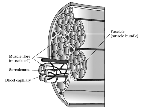
Diagrammatic cross sectional view of a muscle showing muscle bundles and muscle fibres - A few muscle fibres are bundled to form a fascicle
- A fascicle is surrounded by a connective tissue sheath called Perimysium
- All the fascicles are surrounded by a common collagenous tough sheath of connective tissue layer called ‘fascia’
- The whole muscle is surrounded by an outer sheath of connective tissue the epimysium
- A skeletal muscle is generally connected to the skeleton by tendon
4. STRUCTURE OF A MUSCLE FIBRE
- A muscle fibre is surrounded by a sarcolemma
- Sarcolemma is a fused structure of basement membrane and plasmalemma
- Living substance inside the sarcolemma is the sarcoplasm

Structure of Muscle fibre - Sarcoplasm has several nuclei
- A cell with many nuclei is multinucleated or syncytial.
- Sarcoplasmic reticulum of a muscle fibre stores calcium ions in the cisternae.
- Many mitochondria or sarcosomes are present in the muscle fibre
- The respiratory pigment of muscle fibre is the
- The parallelly arranged contractile filaments of a muscle fibre are the myofibrils
- A myofibril consists of alternate dark and light bands
- The light band has the contractile protein actin
- The light band is the isotropic band or I-band
- The dark band has the contractile proteins myosin actin
- The dark band is the Anisotropic band or A-band
- The contractile proteins are arranged as rod like structures parallel to each other and also to the longitudinal axis of the myofibrils.
- Actin filaments are the thin filaments
- Myosin filaments are the thick filaments
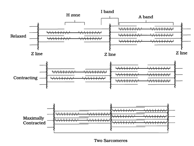
- In the centre of each I-band, there is an elastic fibre called “Z” line or Krause’s membrane or Dobie’sline.
- The thin filaments are attached to the ‘Z’ line.
- The thick filaments in the A band are also held together by a thin fibrous membrane the M-line
- The A-band and I bands are alternately arranged throughout the length of the myofibrils
- The region of the myofibril between two successive ‘Z’ lines is called ‘Sarcomere’
- Sarcomere is the functional unit of contraction
- In a sarcomere, the central part of the A-band has only thick filaments. This is the ‘H’ zone or Hensen’s disc
- Partially overlapped part of the A-band by thin filaments on either side is more darker part of A- band
- I-band has only the thin filaments
- Each sarcomere has one full A-band and two half I-bands.
- Sarcolemma invaginates at some places to form T-tubules(transverse tubules)
- T-tubules help in the conduction of depolarisation
5. STRUCTURE OF CONTRACTILE PROTEINS
- Each thin filament is made of the proteins such as
- F (filamentous) actin, tropomyosin and troponin.
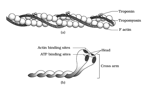
(a) An actin (thin) filament (b) Myosin monomer (Meromyosin)
- Two filamentous (F) actins are helically coiled to each other
- Each “F” actin is a polymer of monomeric globular (G) actin molecules
- Two filaments of tropomyosin run close to the “F” actin molecules throughout their length
- Tropomyosin covers active sites of the actin filaments.
- The active sites of the actin filaments form an actomyosin complex during muscle contraction
- Three-unit-protein called Troponin, binds the tropomyosin.
- Troponin-I binds to actin .Troponin-T binds to tropomyosin. Troponin-C binds to calcium ions at the time of muscle contraction.
- About 200 – 300 monomeric proteins called meromyosin forms a thick filament
- Each meromyosin has two important parts, a globular head with a short arm and a tail
- Globular head and the short arm is formed by heavy meromyosin (H.M.M)
- The tail is formed by the light meromyosin (L.M.M)
- The head and the short arm at regular distances projects outwards to form cross arms
- The myosin binds with the active sites of actin by cross arms
- The head of myosin is an active ATP ase and has a binding site for ATP
Triad System
- Sarcolemma, in many types of muscles, invaginates into the sarcoplasm and forms transverse tubules (T-tubules) at the level of Z line in the non-mammalian vertebrates. In the mammals, they penetrate into the junction between the A and I bands.
- Each T tubule is flanked on either side by several terminal cisternae of the sarcoplasmic reticulum.
- T tubule and the two terminal cisternae at its sides form the triad system. Most of the stored calcium of the sarcocyte is located in the terminal cisternae.
Motor Unit
- A motor neuron and the set of muscle fibres innervated by all the telodendrites constitute a motor unit.
- The junction between a motor neuron and the sarcolemma of a muscle fibre is called the neuromuscular junction or motor end plate.
- Sarcomere of a striated muscle can be considered a ‘functional unit’ of contraction
6. MECHANISM OF MUSCLE CONTRACTION
- Sliding filament theory of Hanson and Huxley explains muscle contraction
- Muscle contraction is initiated by the central nervous system
- Every muscle is innervated by a motor nerve
- The ending of the nerve on the sarcolemma of the muscle is the neuromuscular junction (or) motor end plate.
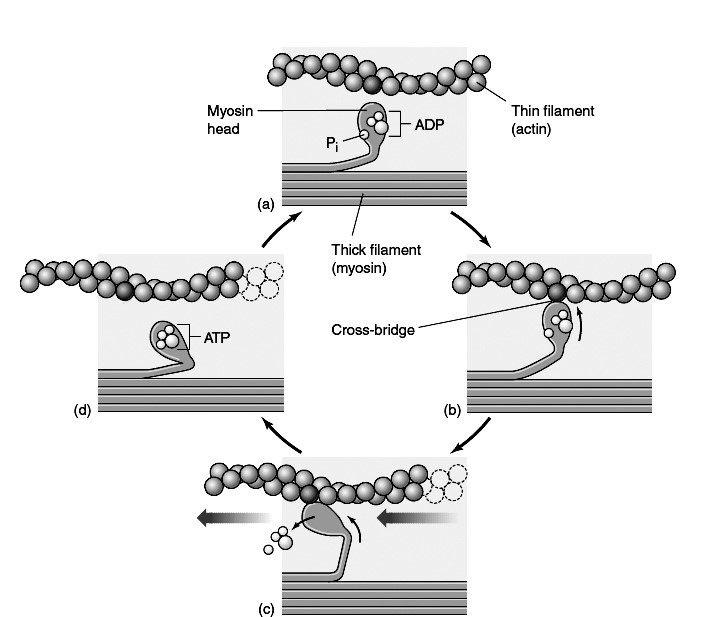
The cross-bridge cycle in muscle contraction. (a) With ADP and Pi attached to the myosin head, (b) the head is in a conformation that canbind to actin and form a crossbridge. (c) Binding causes the myosin head to assume a more bent conformation, moving the thin filament along the thick filament (to the left in this diagram) and releasing ADP and Pi. (d) Binding of ATP to the head detaches the cross-bridge; cleavage of ATP into ADP and Pi puts the head into its original conformation, allowing the cycle to begin again.
- The muscle fibre with its motor neuron forms a motor unit.
- The motor neuron releases a neuro transmitter substance the acetylcholine at motor end plate.
- Acetylcholine depolarises sarcolemma
- The depolarisation reaches the cisternae of the sarcoplasmic reticulum through T-tubules
- Ca++ are liberated into sarcoplasm
- Ca++ binds with Tn-C of Troponin.
- TnC-Ca++ makes the tropomyosin move away from over the active sites of actin. Thus, active sites are exposed.
- Using the energy of ATP by hydrolysis, the myosin head binds to the exposed active site of actin forming an actomyosin complex or cross bridge.
- This cross bridge pulls the actin filament towards the M-line of A-band. This is ‘power stroke’
- Z-line, to which actin filaments are attached, is also pulled towards the M-line of A-band.
- This pulling of successive Z-lines closer to A-band causes the shortening of the sarcomere. This is contraction.
- During contraction, there is no change in the length of A-band.
- The I-bands and H-zone get reduced or may completely disappear.
- The hydrolysis of ATP forms ADP and Pi. ‘Pi’ is released just before the power stroke while ADP is released after power stroke.
- A new ATP binds with the head of the myosin making the cross bridges to detach.
- The actomyosin complex is thus broken, so that the muscle gets back to relaxed state.
- This process continues till the Ca++are pumped back to the cisternae.
- In the absence of Ca++, the active sites are covered by the tropomyosin – This is relaxation.
7. MUSCLE FATIGUE
- Repeated activation of skeletal muscles can lead to the accumulation of lactic acid due to anaerobic break down of glucose in them causing fatigue.
- At this state the skeletal muscle fails to contract temporarily
8. CORI CYCLE
- The lactic acid produced during rapid contraction of skeletal muscles under low availability of O2 is partly oxidised and a major part of it is carried to the liver by the blood.
- In the liver, the lactic acid is converted into pyruvic acid and then to Glucose through Glucogenogenesis
- The Glucose can enter the blood and be carried to muscles and immediately used.
- If the muscle have stopped contraction by the time the Glucose reaches the muscle, the Glucose can be used to rebuild reserve of Glycogen through Glycogenesis.
- The two way traffic between skeletal muscle and liver is called coricycle
9. TYPES OF MUSCLE FIBERS
- Myoglobin content is high in some muscles which are called ‘red muscle fibres’
- Red muscles are having more mitochondria.
- Mitochondria utilise oxygen stored in the myoglobin to produce ATP
- More amount of glycogen is also present in muscle
- Red muscles are also called aerobic muscles
- Some muscles are having less amount of myoglobin. These are called white muscle fibres
- White muscle has less number of mitochondria
- White muscle has more sarcoplasmic reticulum
- Source of energy for white muscle is anaerobic respiration.
10. SKELETAL SYSTEM
- Skeletal system of adult human beings is made up of 206 bones and few cartilages
- Human endoskeleton is grouped into two divisions the axial skeleton and appendicular skeleton.
Axial Skeleton
- It is present along the main axis of the body
- It is made up of 80 bones
- It constitutes the Skull, Vertebral, Column, Sternum and ribs
Skull (22 bones)
- It consists of two sets of bones namely Cranial and Facial bones
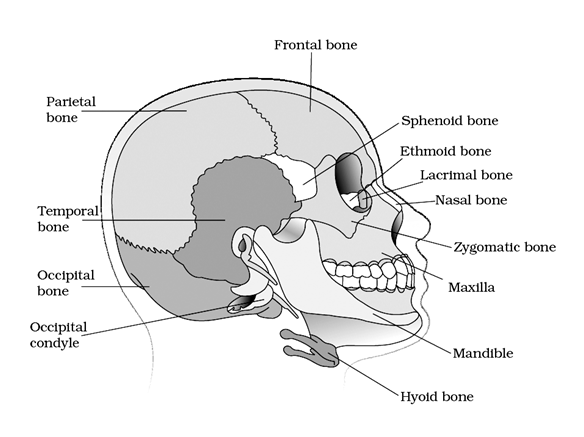
Diagrammatic view of human skull
A. Cranial bones (8)
|
Name of the Bone |
Number of bones |
Location |
| 1) Frontal bone | One | Fore head, anterior part of the Cranial floor and roof of the orbits |
| 2) Parietals | Two | Major portion of the sides and roof of the Cranial Cavity. They are joined to the frontal bone by a coronal suture and posteriorly to the occiput by lambdoid suture |
| 3) Temporal bones | Two | Lateral parts and floor of the cranium |
| 4) Occipital bone | One | Posterior part and most of the base of the cranium |
| 5) Sphenoid bone | One | Present at the middle part of the base of the skull key stone bone |
| 6) Ethmoid bone | One | Present on the middle line of the anterior part of the Cranial floor |
B. Facial Bones (14)
| Bone | Number | Location |
| Nasals | Two | Bridge of the nose |
| M axillae | Two | Form the upper Jaw |
| Zygomatic bones | Two | Cheek bones |
| Lacrimal bones | Two | Smallest bones of the face of the hard palate |
| Nasal conchae | Three pairs (superior, middle and inferior) | Lateral wall of the nasal cavity |
| Vomer | One | Present on the floor of the nasal cavity |
| Mandible | One | Lower Jaw longest and strongest of the facial bones |
C. Skeletal Structures associated with sense organs
- Nasal septum : It divides the nasal cavity into right and left cavities
- Orbits : Bony depressions which accommodate the eye balls and associated structures
- Ear Ossicles : Middle ear contains three tiny bones called
i) Malleus – Modified articular
ii) Incus – Modified quadrate
iii) Stapes – Modified hyomandibular
D. Hyoid bone : It is present at the base of the buccal cavity between the larynx and the mandible.
Vertebral Column (26 vertebrae) :
Structure of Vertebra
1) A vertebra consists of the body (centrum) vertebral arch and many processes for articulation or attachment of muscles
2) Vertebral arch encloses the neural canal through which the spinal cord passes.
3) Articulations between the successive veritebral are called inter vertebral discs.
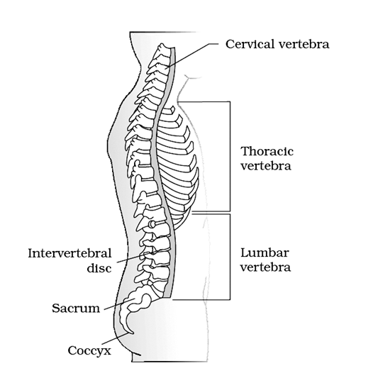
Vertebrae in the Vertebral Column
A) Cervical Vertebrae (7)
i) First vertebra is the Atlas
ii) Second vertebra is the Axis
iii) Atlas articulates with the occipital condyles of the skull
iv) It has an odontoid canal
v) Axis has a strong odontoid process that fits into odontoid canal of the atlas
B) Thoracic Vertebrae (12)
i) The heads of ribs namely capitulum and tuberculum articulates with the articular facets of the thoracic vertebrae
C) Lumbar Vertebrae : Largest and strongest of the vertebrae provide surface for the attachment of the large back muscles
D) Sacrum : Triangular bone formed by the fusion of the five sacral vertebrae.
E) Coccyx : Triangular bone formed by the fusion of four coccygeal vertebrae.
Ribs
- Ribs of human beings are 12 pairs
- Rib is a thin curved flat bone
- Ribs articulate dorsally with the vertebral column
- Capitulum of rib(1st head of rib) articulates with the centrum of the vertebra.
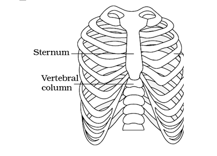
Ribs and rib cage
- Tuberculum of rib(2nd head of rib) articulates with the transverse process of the vertebra
- The ribs with two articular surfaces or heads are called bicephalic ribs or double-headed ribs
- The first 7 pairs of ribs are called true ribs
- True ribs are ventrally connected to the sternum with hyaline cartilage.
- The 8th ,9th and 10th pair of ribs are called vertebrochondral ribs (or) false ribs
- False ribs do not articulate with the sternum
- False ribs join the seventh pair of ribs with hyaline cartilage
- Last two pairs (the 11th and 12th pair) are not connected ventrally. They are called floating ribs
- Ribs and sternum form the rib cage or thoracic cage
- Rib cage protects the heart, lungs and also helps in respiration
Sternum
- The endoskeleton in mid ventral line of thorax is the sternum.
- Sternum of human beings is about 15 cm length
- Sternum is sword – shaped or dagger shaped structure
- Sternum has anterior manubrium, middle body and posterior xiphoid process
Appendicular skeleton
- Endoskeleton attached at an angle to the axial skeleton is the appendicular skeleton
- Skeleton that provides support and articulation of limbs is the appendicular skeleton
- Pectoral girdle, pelvic girdle, bones of forelimbs, bones of hind limbs form the appendicular skeleton.
- Appendicular skeleton of man has 126 bones
Pectoral girdle
- Bony complex supporting and connecting the limbs and axial skeleton is the pectoral girdle
- Pectoral girdle has two halves with Scapulae – 2 and Clavicles – 2
- Clavicle and Scapula are the two bones in each half of the pectoral girdle.
- “S”- shaped, double-curved, thin and elongated bone is the clavicle or the collar bone
- At one end, clavicle is attached to the acromion process of scapula and at its other end with the manubrium of the sternum
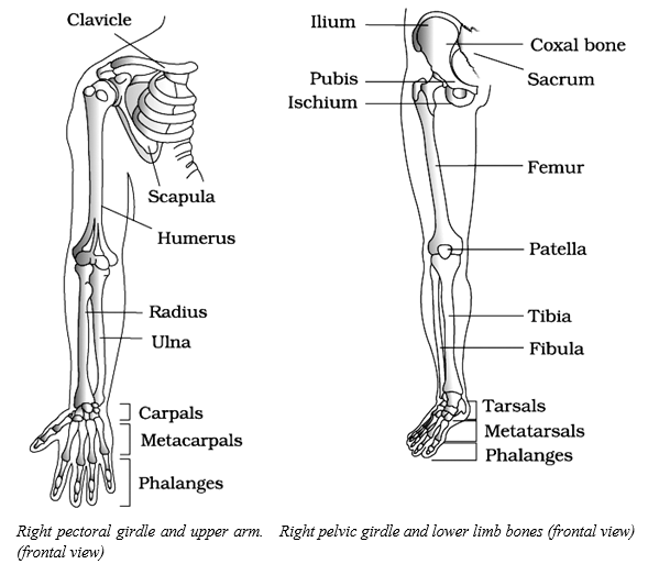
- Thin, flat, triangular curved bone is the scapula – the shoulder blade.
- Scapula is extended on the back side of the thorax between 2nd to 7th
- The hook – like structure of scapula is called coracoid process.
- The diagonally placed sharp protuberance of the scapula is the acromion spine.
- The free end of acromion spine has a thick acromion process
- The glenoid cavity of scapula is for the articulation of the head of the humerus to form a ball and socket joint called shoulder joint.
Pelvic girdle
- The endoskeleton providing articulation of hind limbs is the pelvic girdle
- Pelvic girdle has two coxal bones (innominates)
- Each coxal bone( innominatum) is formed by the fusion of three bones such as Ilium, Ischium and Pubis.
- The cup-shaped cavity formed at the place of the fusion of the three bones is the acetabulum.
- The head of the femur(thigh bone) articulates with the acetabulum to form a ball and socket joint called hip joint.
- The two halves of the pelvic girdle fuse midventrally to form a pubic symphysis
- Pubic symphysis contains fibrous cartilage.
- A large oval gap called obturator foramen is present between pubis and ischium.
Bones of fore limb
- Upper arm – Humerus – 1 ; Fore arm – Radius & Ulna – 2; Wrist bones (carpals) – 8 ; Palmbones ( metacarpals) – 5; Bones of digits (Phalanges) – 14; Total = 30 bones. Both the fore limbs totally have 60 bones
Bones of hind limb
- Thigh bone(Femur) – 1; Shank bones( Tibia & Fibula) – 2 ; Ankle bones (Tarsals) – 7 ; Bones of instep (metatarsals) – 5 ; Bones of digits ( Phalanges) – 14 ; Bones of Knee(Patella)- 1;
- Total bones of each hind limb – 30. Both hind limbs contain a total of 60 bones
11. JOINTS
- This cross bridge pulls the actin filament towards
- Joints are points of contact between bones or
- between bones and cartilages
- Force generated by the muscle is used to
- carry out movement through Joints
Types of Joints
- Joints are classified into three major structural forms namely
(i) Fibrous joints (ii) cartilaginous joints (iii) synovial Joints
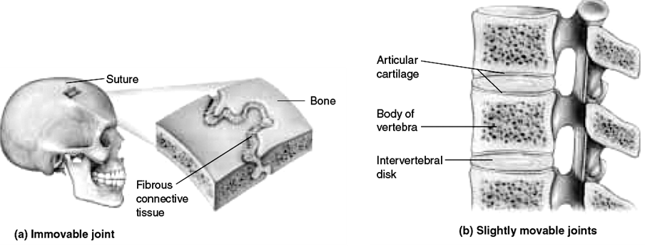
(i) Fibrous Joints
- These are composed of dense irregular connective tissue
- They do not allow any movement
- These are three types
(i) Sutures : Present between the cranial bones. Coronal suture is present between. Frontal and parietal bones.
(ii) Syndesmoses : Interosseous membrane present between tibia and fibula.
(iii) Gomphoses : Dento alveolar Joint - Sutures and gomphoses are synarthoses
- Syndesmose is amphiar those
(ii) Cartilaginous Joints
- Bones are joined together by cartilages
- These are two types
(a) Synchondroses: Epiphyseal plate present between Epiphysis and diaphysis
(b) Symphysis : Pubic symphysis of pelvic girdle. - Synchondrose is a synarthrose and symphysis is an amphiarthose
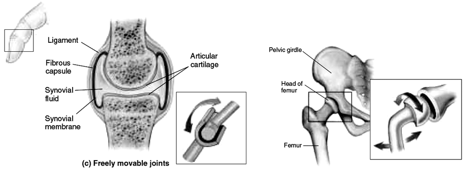
Synovial Joints
- Fluid filled synovial cavity is present between the articular surfaces of two bones
- The articular surfaces of bones are covered by hyaline cartilage
- All synovial Joints are diarthroses
Structure of Synovial Joint
- It is Covered by a double layered synovial capsule.
- The outer layer consists of dense fibrous irregular connective tissue with more collagen fibres
- The outer layer is continuous with the periosteum and resists stretching and prevents the dislocation of Joints
- Some fibres in these membranes are arranged in bundles called ligaments
- The inner layer of synovial capsule is formed of areolar tissue and elastic fibres
- The inner layer secretes a viscous synovial fluid which contains hyaluronic acid and phagocytes
Types of synovial Joints
| Type of Joint | Example |
| a) Ball and socket Joint | Hip Joint–between Femur and pelvic girdle Shoulder Joint–between Humerus and Pectoralgirdle |
| b) Hinge joint | Elbow Joint, knee Joint |
| c) Pivot Joint | Atlanto –axial Joint |
| d) Gliding joint | Intercarpal Joints &Inter tarsal Joints |
| e) Condyloid Joint | Joints between the carpals and metacarpals, joint between occipital condyles and atlas |
| f) Saddle Joint | Joint between the carpal and Metacarpal of the thumb |
12. DISORDERS OF MUSCULAR AND SKELETAL SYSTEMS
- Myasthenia gravis : An autoimmune disorder affecting the neuromuscular junctions leading to fatigue, weakening and paralysis of skeletal muscles. Acetylecholine receptors on the sarcolemma are blocked by antibodies/auto immune bodies, leading to weakness of muscles.
- Muscular dystrophy : Progressive degeneration / wasting of skeletal muscle due to certain genetic disorders such as DMD or nutritional disorders (due to poor reception of stimuli).
- Tetany: Rapid muscle spasms (wild contractions) in muscles repeated prolonged contraction of muscles, caused by low blood calcium level, arising from vitamin D deficiency or underactive parathyroid glands (hypoparathyroidism).
- Arthritis: Inflammation of joints.
- Osteoporosis : Age-related disorder characterized by decreased bone mass (loss of calcium due to reabsorption) whereby chances of fracture increase. A decreased level of estrogen is a common cause, especially in post menopause women.
- Gout : Inflammation of joints due to accumulation of uric acid crystals.