


1. INTRODUCTION
- Cells continually use oxygen for the metabolic reactions that release energy from nutrient molecules and produce ATP. At the same time, these reactions release carbon dioxide. Since an excessive amount of carbon dioxide produces acidity that is toxic to the cells, the excess carbon dioxide must be eliminated quickly and efficiently. It is, therefore evident that oxygen has to be continuously provided to the cells and carbon dioxide produced by the cells has to be released out.
- This process of exchange of O2 from the atmosphere with CO2 produced by cells is called breathing, commonly called Respiration.
2. TYPES OF RESPIRATION
Respiration is of two types depending upon presence and absence of O2.
Aerobic respiration: It occurs in the presence of molecular oxygen. The oxygen completely oxidizes the food into CO2 and water, releasing a large amount of energy.
C6 H12O6 + 6H 2O + 6O2 → 6CO2 + 12H2O + 36 ATP
Anaerobic respiration: It occurs in the absence of molecular oxygen and is also called fermentation.
C6 H12 O6 + 2CO2 → 2C2 H5 OH + 2ATP
Respiration in humans involves the following steps
A) Breathing or Pulmonary ventilation by which atmospheric air with 21% of O2 is drawn and alveolar air rich in CO2 is send out.
B) Diffusion of gases across the alveolar membrane
C) Transport of gases between the lungs and tissue cells.
D) Diffusion of gases between systemic capillaries and the tissues.
E) Utilization of O2 by the cells for catabolic reactions and resultant production H2O and ATP (cellular respiration)
3. IMPORTANT RESPIRATORY ORGANS
- Protozoans, sponges and cnidarians – Exchange of gases occurs through body surface.
- Platyhelminthes and Nemathelminthes – No distinctive organs of respiration.
- Free living animals – through body surface Parasites – No exchange of gases (Anaerobic respiration)
- Annelids – Moist skin- Cutaneous respiration in earthworms and Parapodia in some Polychaetes.
- Arthropods – Limulus-book gills. Arachnids-book lungs and/ or trachea
- Crustaceans – Gills – Insects, millipedes and centipedes – Tracheae
- Molluscs – Ctenidia (gills) and pulmonary sac.
- Echinodermata – Tube feet in all, Dermal branchiae- sea stars, Genital bursae- brittle stars, Peristomial gills-sea urchins, Respiratory trees-sea cucumbers.
- Hemichordata – Pharyngeal wall
- Urochordates – Pharyngeal wall
- Cephalochordata– Body wall
- Vertebrates – Cyclostomes, Fishes – Gills
- Amphibians –Skin,Lungs and buccopharyngeal lining.
- Reptiles, Birds, Mammals – Lungs
4. HUMAN RESPIRATORY SYSTEM
Human respiratory system is divided into two parts
1) Upper respiratory tract: It includes Nose and Pharynx
2) Lower respiratory tract: It includes Larynx, Trachea, Bronchi, Lungs
Nose:
- Nose is the outer most part of respiratory tract, it surrounds nasal cavity.
- It opens to the outside with two openings called nostrils or external nares, internally (posteriorly) opens in pharynx through two internal nares (Choanae)
- The floor of the nasal cavity, which forms the roof of the mouth, is made up by the bones of the hard palate
- Nasal cavity is divided into two nasal chambers by a vertical partition nasal septum
- Each nasal chamber is divided into three parts
- Vestibular part
- Respiratory part
- Olfactory part
- Vestibule It is the anterior part of the nasal chamber, which has hair and sebaceous glands to prevent the entry of dust particles.
- Respiratory part : It is the middle large part. Its lateral walls have three thin twisted bones called superior, middle and inferior conchae or turbinals. This region functions as air conditioner, it brings the temperature of the incoming air on par with the body temperature.
- Olfactory part The superior part of the nasal cavity has olfactory region. It helps in olfaction.
Pharynx
- It is 13 cm long, starting with soft palate area representing the crossing point of air route and food route.
- It has three parts Nasopharynx, Oropharynx, and Laryngopharynx.
- Uvula, the free posterior and muscular part of soft palate which divides pharynx as anterior oropharynx and posterior nasopharynx.
- Nasopharynx is the upper part of the pharynx behind nasal cavity. There are four openings in its walls, two internal nares and two openings of eustachian tubes.
- Oropharynx is the middle part, which is a continuation of buccal cavity.
- Lower part of the pharynx is laryngopharynx opens into the oesophagus through gullet and larynx through glottis anteriorly.
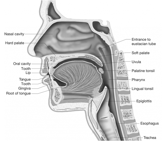
Larynx (Voice box)
- The upper part of trachea is made up of cartilaginous box called larynx.
- Larynx is made up of nine cartilages three of them are unpaired while three of them are paired cartilages.
- The unpaired cartilages are Thyroid, Cricoid and Epiglottis.
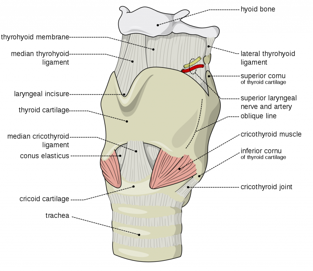
- Thyroid cartilage It is the largest cartilage of C-shaped incomplete dorsally. In males it is larger and protrude in the neck area where it is called Adam’s apple.
- On anterior part of thyroid, epiglottis is attached which is composed of yellow elastic cartilage and covers the glottis like a flap.
- Cricoid is a ring shaped cartilage and constitute the inner part of the larynx
- The paired cartilages are Arytenoid, Cuneiform and Corniculate.
- Arytenoid is pyramid shaped, composed of hyaline cartilage and forms the dorsal side of larynx.
- Between thyroid and arytenoid elastic fibres are present. These are called vocal cords.
- The anterior folds above the vocal cords are called false vocal cords.
- The gap between the vocal cords is called rimaglotidis /peep hole
- Corniculate: These are two conical nodules of elastic fibro-cartilage, which lie at the apices of arytenoid cartilages.
- Cuneiform: There are two small and elongated club shaped nodules of elastic fibro-cartilage, which lie above and anterior to corniculate cartilages.
- When air is forced through the larynx, it causes vibration of the true vocal cords and sound is produced.
- The pitch of a sound is determined by the tension on the vocal cords – the greater the tension, the higher the pitch.
- Speech occurs during expiration when the tongue, cheeks and lips manipulate the sounds produced by the vocal cords
- In human male the vocal cords are longer and thicker under the influence of androgens. This changes the male sound as deep or low pitch at puberty.
- In children and female vocal cords are usually short (as no significant thickening occurs in the female at puberty) and the voice is sharp or high pitch.
Trachea (wind pipe)
- It is a straight tube located anterior to oesophagus. It extends from larynx to midthoracic cavity.
- It is supported by incomplete C-shaped rings of hyaline cartilage and lined by Pseudostratified ciliated epithelium.
- All the cilia of trachea beat towards pharynx and push the mucus along with it. This ciliary movement helps in the cleansing of lungs.
- At the level of 5th thoracic vertebra trachea divides into right and left primary bronchi. These bronchi enter the lungs of their side
Bronchi
- Inside the lungs bronchi keep on dividing .During this division the cartilage rings become smaller and irregular.
- Primary bronchi divides to form secondary bronchi, tertiary brochi and terminal bronchi.
- All bronchi are lined by ciliated pseudostratified epithelium and supported by incomplete cartilaginous rings.
- The tertiary bronchi divide forming primary, secondary, tertiary, terminal bronchioles and respiratory bronchioles
- Each respiratory bronchiole forms an alveolar duct and alveolar sacs
- An alveolar sac ends in vascularized bags called alveoli or air sacs.
- The system of branching and rebranching of the respiratory passageways is called respiratory tree or bronchial tree.
- Later bronchioles are without cartilaginous rings. They are lined by ciliated cuboidal or columnar epithelium.
Lungs
- These are paired, cone shaped, spongy organs in the thoracic cavity enclosed within pleural cavity.
- The outer pleura attached to the wall of the thoracic cavity is called parietal pleura and the inner layer is called the visceral pleura
- In between both the membranes a very narrow cavity called Pleural cavity is present.
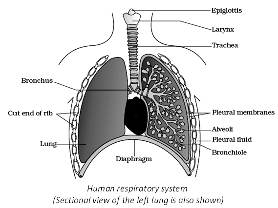
- In this cavity, a very thin layer of pleural fluid is present to reduce the friction and makes the movement of lung easy.
- The part starting with external nostrils up to the terminal bronchioles constitute the conducting part
- The alveoli and their ducts form respiratory or exchange part
- The right and left lungs are divided into three and two lobes respectively.
- Space between right and left pleural cavities is mediastinum and left lung contains a concavity called the cardiac notch in which lies the heart.
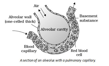
- Exchange of respiratory gases takes place by simple diffusion across the alveoli and capillary walls. Lungs contain about 300 million alveoli with surface area of 70 m2.
- Branches of pulmonary artery supply deoxygenated blood to each lung.
- Respiratory gases are exchanged between the blood and alveolar air by diffusion through alveolar wall.
- Pulmonary veins leave the lungs and convey oxygenated blood to the left atrium of the heart.
5. HUMAN RESPIRATORY SYSTEM
- Respiratory membrane is 0.5 m in thickness. Alveolar epithelium and its basement membrane, thin interstitial space and capillary basement membrane and endothelial membrane. Alveoli are functional units of lungs. Three types of cells line them
1) Type I Alveolar cells/ pneumocytes
2) Type II Alveolar cells/ pneumocytes
3) Dust cells or Alveolar macrophages - Type I Alveolar cells participate in exchange of gases and associate with the endothelial membrane of pulmonary capillaries and form 5. Respiratory membrane through which exchange of gases takes place.
- Type II Alveolar cells secrete surfactant. 6. Surfactant is a mixture of mucus and phospholipids and reduces the surface tension during exchange of gases, which reduces the tendency of alveoli to collapse.
- Dust cells or Alveolar macrophages- wandering macrophages ingesting fine dust particles and other debris.
6. MECHANISM OF BREATHING
- It takes place by the changes in volume of airtight thoracic cavity,
- Lower boundary of thoracic cavity is formed by diaphragm which divides body cavity into upper thoracic and lower abdominal cavity.
- Diaphragm is an elastic, dome-shaped, plate like structure with its convex side towards thoracic cavity
- 12 pair of ribs obliquely attached between vertebral column and movable sternum form the thoracic cage.
- Last two pairs of ribs are floating ribs, which have no role in breathing.
- Two sets of muscles, external intercostal muscles and internal intercostal muscles are present between the ribs.
In humans breathing involves two processes
1. Inspiration
2. Expiration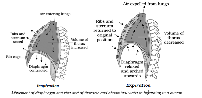
Inspiration
- It takes place by the contraction of phrenic (radial) muscles, which pulls the diaphragm downwards increasing the volume of thoracic cavity.
- Abdominal muscles relax and allow compression of abdominal organs by the diaphragm contraction of external intercostal muscles lift up the ribs and sternum, hence volume of thoracic cavity increases towards this side.(dorso-ventral axis)
- This increase in volume and decreases the pressure in thoracic cavity so the air enters into the lungs.
- The elevation of thoracic cavity is supported by external intercostal muscles therefore these muscles are defined as inspiratory muscles.
Expiration
- During expiration diaphragm and external intercostal muscles simply relaxes which reduces the thoracic volume there by pulmonary volume.
- This leads to increase in intra pulmonary pressure to slightly above that of atmospheric pressure, causing the expulsion of air form the lungs.
- In humans expiration is a passive process and no muscles are defined as expiratory muscles during normal quiet expiration.
- But during forceful expiration internal intercostal and abdominal muscles participate and called expiratory muscles.
- On an average, a healthy human breaths 12- 16times/minute.
- The volume of the air involved in breathing movements can be estimated by using a spirometer. The record is called spirogram
7. RESPIRATORY VOLUMES
- Tidal Volume (TV): It is the volume of air inspired or expired during normal inspiration or expiration. It is approximately 500 ml.
- Inspiratory Reserve Volume (IRV): It is the additional volume of air that can be inspired forcefully after the end of normal inspiration. It is 2500-3000 ml.
- Expiratory Reserve Volume (ERV): It is the additional volume of air that can be expired our forcefully after the normal expiration. It is 1000-1100 ml.
- Residual Volume (RV): It is the volume of air remaining in the lungs even after forceful expiration. It is 1100-1200 ml.
Lung capacities
- Inspiratory Capacity (IC): It is the total volume of air that a person can inhale after normal expiration. IC = TV + IRV. It is about 3000-3500 ml.
- Functional Residual Capacity (FRC): It is volume of air remaining in the lungs after normal expiration. FRC = ERV + RV.
- Vital capacity (VC): It is the maximum volume of air that can be expelled out forcefully after a deep or maximal inspiration. VC = IRV + TV + ERV.
- Total Lung Capacity (TLC): It is the volume of air present in the lungs after a deep or maximal inspiration. TLC = IRV + TV + ERV + RV.
8. EXCHANGE OF GASES
- It takes place in the alveoli by simple diffusion due to difference of partial pressure of CO2 and O2 in the alveoli and the perialveolar blood capillaries.
- The rate of gas exchange depends on partial pressure of the gases, solubility of the gases, surface area and diffusion distance.
- A gas always diffuses from the region of its higher partial pressure to the region of its lower pressure.
- As the diffusing capacity of CO2 is 20 – 25 times higher than that of O2. CO2 of blood rapidly passes out into alveolar air
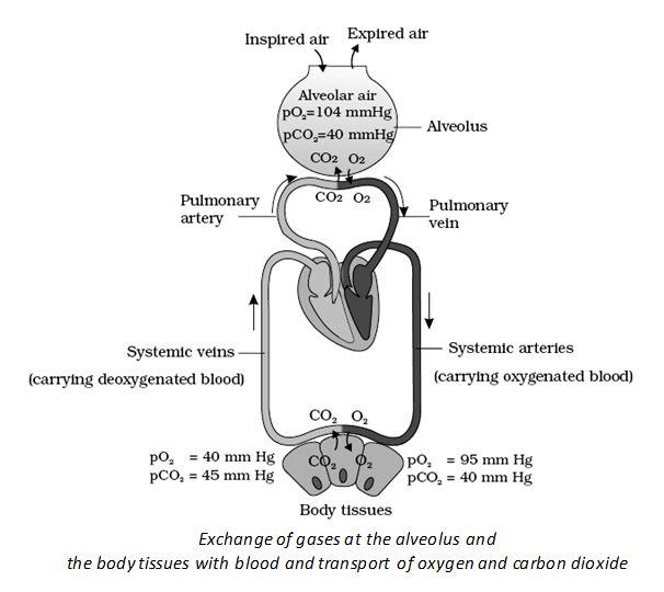
| Partial Pressures (in mm Hg) of Oxygen and Carbon dioxide at Different Parts Involved in Diffusion in Comparison to those in Atmosphere | |||||
| Respiratory Gas | Atmospheric Air | Alveoli | Blood (Deoxygenated) | Blood (Oxygenated) | Tissues |
| O2 | 159 | 104 | 40 | 95 | 40 |
| CO2 | 0.3 | 40 | 45 | 40 | 45 |
9. TRANSPORT OF GASES
- Blood is the medium to carry O2 from lungs to tissue and CO2 from tissue to lungs.
Transport of Oxygen
(i) Transport of oxygen through plasma: Being less soluble in water, only 3% of O2 is transported as dissolved form in plasma, this is only a negligible amount.
(ii) Transport of oxygen by RBC: 97% is transported in bound form through the respiratory pigment, haemoglobin (Hb).
- Haemoglobin is a respiratory pigment in the blood of vertebrates is Haemoglobin consists of 4 Polypeptide chains each with 1 haem group as non-protein part. 1 Fe2+ is at the centre of each polypeptide chain.
- 1 Fe atom combines with 1 molecule of O2
- 1 molecule of Hb combines with 4 molecule of O2
- Combining with O2 it forms oxyhaemoglobin, an unstable compound that dissociates at lower pressure of O2 in the tissue undergoing metabolism.
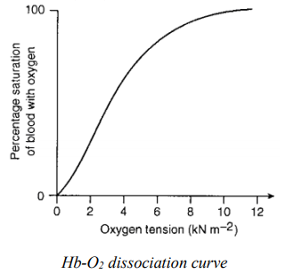
- It explains the relationship between percentage saturation of haemoglobin and partial pressure of oxygen. The Hb-O2 dissociation curve is typically a sigmoid curve.
- The saturation of Hb at 100 mm Hg pO2 is 97%
- Saturation of Hb at 40 mm Hg pO2 is 75%. It means only 22% (1/5th) of oxygen is unloaded
- in the tissues. The remaining 4/5th is in the form of reserve in the blood itself
- If he skeletal muscles are involved in vigorous exercise more oxygen is given away rapidly (up to 62% at 20mm Hg of pO2)
- In a resting person Hb always carries 70% of oxygen, this ensures supply of oxygen for survival for 4 – 5 minutes after stopping of heart or when breathing is interrupted.
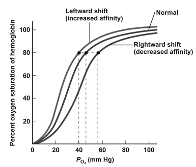
Factors affecting Hb-O2 dissociation curve
- Effect of pCO2 : If the pCO2 level is higher at the site of metabolism (tissue) the Hb releases O2 at faster rate hence the curve shifts to right hand side.
- Effect of pH: Lower pH causes dissociation of So, the curve shifts to right hand side.
- Effect of Temperature: If the temperature is higher at the site of metabolism, the Hb releases oxygen at faster rate, hence the curve shifts to right hand side.
Shifting of oxyhaemogobin –dissociation curve
- In above conditions the dissociation curve shifts to right hand side and the affinity of Hb for oxygen decreases to release more O2 to the tissues.
- Higher pH, lower CO2 and lower temperature shifts the curve to the left side.
- The effect of PCO2 and H+ concentration on oxygen affinity of haemoglobin is called Bohr effect.
- Raise in PCO2 and fall in pH decrease the affinity of haemoglobin for oxygen on the other hand a fall in PCO2 and raise in pH increases affinity of haemoglobin for oxygen
Transport of Carbon dioxide
CO2 is highly soluble in water and hence transported in following three ways
(i) In dissolved form : 7% of CO2 is carried in a dissolved state (physical solution) through plasma
CO2 + H 2O → H2 CO3
(ii) As carbamino compounds : About 20 – 25 % of CO2 is transported by combining with amino group of plasma proteins or Hb. Hb + NH2 + CO2 → Hb NHCOO H
- Hb is an amphoteric compound. It has the peculiar character of taking more oxygen in O2 rich areas and leave CO2. It accepts more CO2 in areas where the CO2 is more and leave oxygen.
(iii) As bicarbonates: About 70% of CO2 is transported as bicarbonates.
- CO2 diffuses into the RBC at tissues as PCO2 is more in tissues.
- CO2 combines with H2O and form carbonic acid under the presence of carbonic anhydrase. CO2 + H 2O → H2CO3
- Carbonic acid dissociates into HCO3 and H+
- HCO3 diffuse into the blood plasma from the RBC. The H+ ions are buffered by Hb which turns into HHb. As a result O2 is released.
- To maintain electrical balance Cl ions diffuse from plasma into RBC this is called chloride shift or Hamburger’s phenomenon.
- In pulmonary capillaries high PO2 causes removal of H+ from Hb. Released H+ combines with HCO and form H2 CO3
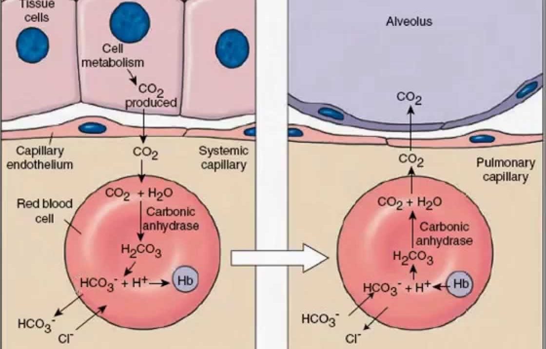
- Due to the decrease of HCO3 concentration in RBC, HCO from plasma moves into the RBC
- and Cl moves out this is reverse chloride shift.
- H2CO3 dissociation and gives rise to H2O and CO2 and CO2 diffuses into the alveoli.
H 2 CO3 → H 2O + CO2
10. REGULATION OF RESPIRATORY MOVEMENTS
- Human beings can maintain and moderate the respiratory rhythm to suit the demands of body tissues.
- ‘Respiratory rhythm centre’ present in medulla oblongata is responsible for this regulation.
- ‘Pneumotaxic centre’ present in pons can moderated the functions of Respiratory rhythm centre.
- Neural signal from Pneumotaxic centre can reduce the duration of inspiration and there by alter the respiration rate.
- A ‘Chemosensitive area’ is situated adjacent to ‘Respiratory rhythm centre’ and is highly sensitive to CO2 and H+
- Increase in CO2 and H+ ions activate ‘chemosensitive area’ which in turn can send signals to ‘Respiratory rhythm centre’ which makes necessary adjustments in the respiratory process by which these substances are eliminated.
- Receptors in Aortic arch and Carotid artery also recognise changes in CO2 and H+ ion concentration and send signal to ‘Respiratory rhythm centre’ which makes necessary adjustments to bring their amounts to normal.
- The role of Oxygen in the regulation of respiratory rhythm is quite insignificant.
11. DISORDERS OF RESPIRATORY SYSTEM
1. Asthma
- It is a difficulty in breathing caused due to the inflammation of bronchi and bronchioles.
- Allergens cause release of histamine and other inflammatory substances which causes constriction of the bronchi.
- Symptoms include difficulty in breathing and wheezing.
2. Emphysema
- It is a chronic disorder in which alveolar walls are damaged and their walls coaleasce due to which respiratory surface area decreases.
- The lung shows larger but fewer alveoli and more fibrous and less elastic.
- The major cause of this is smoking of tobacco.
3. Bronchitis
- Inflammation of bronchi resulting in the swelling of mucous lining of bronchi.
- Symptoms include chronic cough with thick mucus / sputum (phlegm)
4. Pneumonia
- It is the infection of lungs caused by bacteria such as Streptococcus pneumoniae
- It can also be caused by certain viruses, fungi, protozoans and mycoplasma
- Symptoms include inflammation of lungs, accumulation of mucus in alveloi and impaired exchange of gases, leading to death.
- Emphysema, bronchitis and asthma come under
- Chronic Obstructive Pulmonary Diseases (COPDs)
5. Occupational Respiratory disorders
- Asbestosis occurs due to chronic exposure to asbestos dust.
- Silicosis occurs because of long term exposure to silica dust
- Siderosis occurs due to deposition of iron particle in tissues.
- Pneumoconicosis, Hyperferremia and Hemosiderosis are the types of siderosis.
- Black lung disease occurs due of the inhalation of coal dust.