1. Introduction to Human Eye
Our eyes enable us to see the beautiful world around us. The most important part of our eyes is a convex lens inside it that is made of living cells.
The human eye is like a camera having a lens on one side and a sensitive screen called the retina on the other.
2. Human Eye parts and Functions
The essential parts of a human eye are shown in figure.
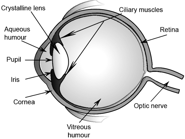
a) Sclerotic: It is the outermost converging of the eye ball. It is made of white tough fibrous tissues. Its function is to house and protect vital internal parts of eye.
b) Cornea: It is the front bulging part of the eye. It is made of transparent tissues. Its function is to act as a window to world. i.e., to allow the light to enter in the eye ball.
c) Choroid: It is a grey membrane attached to the sclerotic from the inner side. Its function is to darken the eye from inside and, hence, prevent any internal reflection.
d) Optic Nerve: It is a bundle of approximately 70,000 nerves originating from brain and entering eye ball from behind. Its function is to carry optical message (visual messages) to the brain.
e) Retina: The optic nerve on entering the ball, spreads like a canopy, such that each nerve and attaches itself to the choroid. The nerve endings form a hemi-spherical screen called retina. These nerve endings on the retina are sensitive to visible light. On the retina are two important areas which we will discuss separately. The function of retina is to receive the optical image of the object and then convert it to optical pulses. These pulses are then sent to the brain through optic nerve.
f) Yellow spot: It is a small area facing the eye lens. It has high concentration of nerve endings and is slightly raised as well as slightly yellow in colour. Its function is to very clear image by sending a large number of optical pulses to brain.
g) Blind spot: It is a region on the retina, where the optic nerve enters the eye ball. It has no nerve endings and hence, is insensitive to the light. It does not seem to have any function. Any image formed on this spot is not visible.
h) Crystalline lens: It is a double convex lens made of transparent tissues. It is held in position by a ring of muscles, commonly called ciliary muscles. Its function is to focus the images of different objects clearly on the retina.
i) Ciliary muscles: It is a ring of muscles which holds the crystalline lens in position. When these muscles relax, they increase the focal length of the crystalline lens and vice versa. Its function is to alter the focal length of crystalline lens so that the images of the objects, situated at different distances, are clearly focussed on the retina.
j) Iris: It is a circular diaphragm suspended in front of the crystalline lens. It has a tiny hole in the middle and is commonly called pupil. It has tiny muscles arranged radially around the pupil. These muscles can increase or decrease the diameter of the pupil. The iris is heavily pigmented. The colour of eyes depends upon colour of pigment. The function of iris is to control the amount of light entering in eye. This is done by increasing or decreasing the diameter of pupil.
k) Vitreous humour: It is a dense jelly like fluid, slightly grey in colour, filling the part of eye between crystalline lens and retina. Its function is (i) to prevent the eye ball from collapsing due to change in atmospheric pressure (ii) in focussing the rays clearly on the retina.
l) Aqueous humour: It is a watery, saline fluid, filling the part of the eye between the cornea and the crystalline lens. Its function is (i) to prevent front part of the eye ball from collapsing with the change in atmospheric pressure (ii) to keep the cornea moist.
3. Power of Accomodation
Have you wondered why the eye is able to focus the images of objects lying at various distances?
It is made possible because the focal length of the human lens can change i.e., increase or decrease, depending on the distance of objects. It is the ciliary muscles that can modify the curvature of the lens to change its focal length.
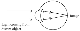
To see a distant object clearly, the focal length of the lens should be larger. For this, the ciliary muscles relax to decrease the curvature and thereby increase the focal length of the lens. Hence, the lens becomes thin. This enables you to see the distant object clearly.
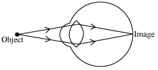
To see the nearby objects clearly, the focal length of the lens should be shorter. For this, the ciliary muscles contract to increase the curvature and thereby decrease the focal length of the lens. Hence, the lens becomes thick. This enables you to see the nearby objects clearly.
The ability of the eye lens to adjust its focal length accordingly as the object distances is called power of accommodation.
The minimum distance of the object by which clear distinct image can be obtained on the retina is called least distance of distinct vision. It is equal to 25 cm for a normal eye. The focal length of the eye lens cannot be decreased below this minimum limit of object distance.
The far point of a normal eye is infinity. It is the farthest point up to which the eye can see objects clearly.
The range of vision of a normal eye is from 25 cm to infinity.
Have you ever thought why animals’ eyes are positioned on their heads?
This is because it provides them with the widest possible field of view. Our eyes are located in front of our face. One eye provides 150° wide field of view while both eyes simultaneously provide 180° wide field of view. It is the importance of the presence of two eyes as both eyes together provide the three-dimensional depth in the image.
4. Defects of Vision
The loss of power of accommodation of an eye results in the defects of vision.
There are three defects of vision called refractive defects. They are myopia, hypermetropia, and presbyopia. In this section, we will learn about these defects of vision in detail.
1. Myopia (short sightedness)
Myopia is a defect of vision in which a person clearly sees all the nearby objects, but is unable to see the distant objects comfortably and his eye is known as a myopic eye.
A myopic eye has its far point nearer than infinity. It forms the image of a distant object in front of its retina as shown in the figure.
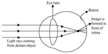
Myopia is caused by
i. increase in curvature of the lens
ii. increase in length of the eyeball
Since a concave lens has an ability to diverge incoming rays, it is used to correct this defect of vision. The image is allowed to form at the retina by using a concave lens of suitable power as shown in the given figure.
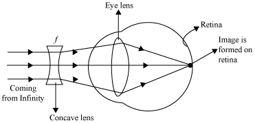
Power of the correcting concave lens
The lens formula can be used to calculate the focal length and hence the power of the myopia correcting lens.
In this case,
Object distance,
Image distance, v = person’s far point
Focal length, f =?
Hence, lens formula becomes
In case of a concave lens, the image is formed in front of the lens i.e., on the same side of the object.
Focal length = – Far point
Now, Power of the required lens
Example: A person can clearly see up to a maximum distance of 100 cm only. Calculate the power of the required lens that can correct his defect?
Solution: Since the person is not able to see farther than 100 cm, he is suffering from myopia. Hence, a concave lens of suitable power is required to correct his defect. The focal length of the lens is given by his far point i.e.,
Focal length = – Far point
= 100 cm
Power of the lens =
Hence, a concave lens of power –1 D is required to correct the given defect of vision.
2. Hypermetropia (Long sightedness)
Hypermetropia is a defect of vision in which a person can see distant objects clearly and distinctively, but is not able to see nearby objects comfortably and clearly.
So, now you can easily represent the problem with a hypermetropic eye with the help of a diagram. It is shown in the given figure.
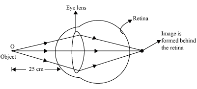
A hypermetropic eye has its least distance of distinct vision greater than 25 cm.
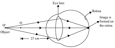 Hypermetropia is caused due to
i. reduction in the curvature of the lens
ii. decrease in the length of the eyeball
Since a convex lens has the ability to converge incoming rays, it can be used to correct this defect of vision, as you already have seen in the animation. The ray diagram for the corrective measure for a hypermetropic eye is shown in the given figure.
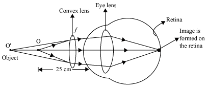
Power of the correcting convex lens
Lens formula, can be used to calculate focal length f and hence power P of the correcting convex lens, where
Object distance, u = –25 cm, normal near point
Image distance, v = defective near point
Hence, the lens formula is reduced to
Example: The defective near point of an eye is 150 cm. Calculate the power of the correcting convex lens that would correct this defect of vision.
Solution: Given that, hypermetropic near point = 150 cm
Hence, image distance, v = – 150 cm
We have the correction formula,
Power of the correcting convex lens,
Hence, a convex lens of power 3.3 D is required to correct the given defect of vision.
3. Presbyopia (Ageing vision defect)
Presbyopia is a common defect of vision, which generally occurs at old age. A person suffering from this type of defect of vision cannot see nearby objects clearly and distinctively. A presbyopic eye has its near point greater than 25 cm and it gradually increases as the eye becomes older.
Presbyopia is caused by the
i. weakening of the ciliary muscles
ii. reduction in the flexibility of the eye lens

i. A person with presbyopia cannot read letters without spectacles. It may also happen that a person suffers from both myopia and hypermetropia. This type of defect can be corrected by using bi-focal lenses. A bifocal lens consists of both convex lens (to correct hypermetropia) and concave lens (to correct myopia).
ii. It is a common misconception among people that the use of spectacles “cures” the defects of vision. However, this is not true as spectacles only “restore” the defects of vision to the normal value.
iii. Cataract
It is also one of the eye defects found commonly in people of older ages. In this defect, the crystalline lens becomes milky and cloudy. This condition is also known as cataract. This causes partial or complete loss of vision. This loss of vision can be restored by removing the cataract by means of a cataract surgery. The use of any kind of spectacle lenses does not provide any help against this defect of vision.
5. Refraction of light through a glass prism
When a ray of light is incident on a rectangular glass slab, after refracting through the slab, it gets displaced laterally. As a result, the emergent ray comes out parallel to the incident ray. Does the same happen if a ray of light passes through a glass prism?
Unlike a rectangular slab, the sides of a glass prism are inclined at an angle called the angle of prism. Therefore, a ray of light incident on its surface, after refraction, will not emerge parallel to the incident light ray (as seen in the case of a rectangular slab).
Refraction of light through a glass prism
To observe the refraction of light through a glass prism, we can perform the following activity.
Take a triangular glass prism, paper sheet, and a few drawing pins. Fix the sheet on a drawing board with the help of drawing pins. Now, place the glass prism on the sheet and draw the outline MNP of the prism on the sheet (as shown in the figure). Draw a straight line AB on the sheet in such a way that it makes some angle with the face MN of the prism. Now, fix two pins on this line and mark them as R and S respectively.
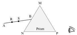
Now, observe the pins R and S through the other side of the prism. Move your head laterally to see the two pins R and S in a straight line. Fix a pin on the sheet near the prism on your side and mark it as T.
Repeat the same step and try to observe the three pins R, S, and T in a straight line. Fix another pin on the sheet so that all four pins appear to be in a straight line when looked through the prism. Draw a straight line CD that passes through the third and the fourth pin i.e., T and W respectively (see figure).
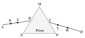
Now, remove the prism and join points B and C. The straight line AB, BC, and CD shows the path of the light ray. It is clear that the path of light is not a straight line since light bends towards the base NP.
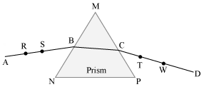
What causes the light to bend when passed through a prism?
Light bends because of refraction that takes place at points B and C respectively, when it tries to enter and emerge from the prism.
Now, draw a straight line normal to side MN and let it pass through point B. Similarly, draw a straight line normal to side MP and let it pass through point C.
Here, line AB = Incident ray
Line BC = Refracted ray
Line CD = Emergent ray
Angle i = Angle of incidence
Angle r = Angle of refraction
Angle e = Angle of emergence
Angle = Angle of deviation
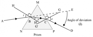
Hence, you will get the path of light ray AB when it travels through a glass prism. The ray AB will bend towards the normal at point B and follow the path BC. Again, it bends away from the normal at C, when it tries to emerge from the prism. This is because the refractive index of air is less than that of glass. Thus, the incident ray AB will not follow a straight line BE.
The extent of deviation of the light ray from its path BE to path CD is known as the angle of deviation ().
6. Dispersion of White Light
Do you know what happens when you take white light as incident ray instead of single ray?
A beam of white light will split into a band of seven colours. The splitting of a beam of white light into its seven constituent colours, when it passes through a glass prism, is called the dispersion of light.
Dispersion of white light by a prism
Isaac Newton was one of the greatest mathematicians and physicists the world ever saw. In 1665, with the help of an experiment he showed that white sunlight is actually a mixture of seven different colours. These constituent colours of white light can be separated with the help of a glass prism.
Take a glass prism and allow a narrow beam of sunlight to fall on one of its rectangular surfaces. You will obtain a coloured spectrum with red and violet colour at its extreme. Try to obtain a sharp coloured band on the screen by slightly rotating the prism. Count the colours of the band and write the sequence of the colours.
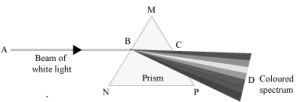
Do you know why white light gets dispersed into seven colours?
When a beam of white light AB enters a prism, it gets refracted at point B and splits into its seven constituent colours, viz. violet, indigo, blue, green, yellow, orange, and red. The acronym for the seven constituent colours of white light is VIBGYOR. This splitting of the light rays occurs because of the different angles of bending for each colour. Hence, each colour while passing through the prism bends at different angles with respect to the incident beam. This gives rise to the formation of the colour spectrum.
Can you say which colour undergoes maximum deviation?
Violet light bends the most whereas red colour deviates least.
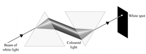
However, Newton did not stop at this point. He thought that if seven colours can be obtained from a white light beam, is it possible to obtain white light back from the seven colours?
For this, he placed an inverted prism in the path of a colour band. He was amazed to see that only a beam of white light comes out from the second prism. It was at this point that Newton concluded that white light comprises of seven component colours.
Formation of a rainbow
The rainbow is a natural phenomenon in which white sunlight splits into beautiful colours by water droplets, which remain suspended in air after the rain.
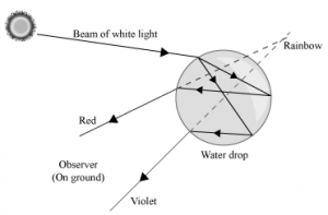
If we stand with our back towards the sun, then we can see the spectrum of these seven colours.
Do you know why a rainbow is shaped similar to an arc?
This is because the rainbow is formed by the dispersion of white light by spherical water droplets. It is the shape of the water droplets that gives the rainbow an arc shape.
A rainbow appears arc-shaped for an observer on ground. However, if he sees the rainbow from an airplane, then he will be able to see a complete circle. This is because he can observe the drops that are above him as well as below him.


