


1. NUTRITION
“Nutrition” is a process of intake as well as utilization of nutrients by an organism. It is the process of breakdown of nutrients into smaller molecules and their absorption. Food provides us nutrition and energy. It contains different types of nutrients in varying amounts according to the need of our body.
Nutrients
These are the substances required by our body for its growth, repair, work and maintenance of the body. Different types of nutrients are carbohydrates, fats, proteins, vitamins, mineral etc. Our daily energy need may vary according to our occupation, age, sex and under some specific conditions.
2. MODES OR NUTRITION
There are several modes of nutrition on the basis of which organisms are classified as follows :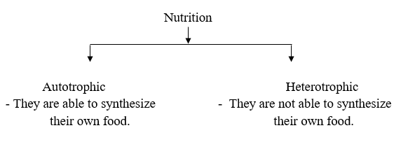
Autotrophic
(Auto = self, trophic = food) It is a mode of nutrition in which organisms prepare their own food. Inorganic molecules like CO2 and H2O are converted into organic molecules like carbohydrates in the presence of sunlight and chlorophyll. e.g. Green plants. Autotrophs are further categorized. as :
(i) Photoautotroph : Those which utilize sunlight for preparing their food
(ii) Chemoautotroph : Those which utilize chemical energy for preparing their food.
Heterotrophic
(Hetero = different ; trophic = food) It is a mode of nutrition in which organisms derive their food from some other animals or plants. They cannot prepare their own food e.g. human being. Heterotrophs are further categorized depending on the nature of food they consume :
(i) Herbivores : Animals which eat only plants, e.g. Cow, goat etc.
(ii) Carnivores : They feed on flesh of other animals, e.g. Lion, vulture etc.
(iii) Omnivores : They feed on plants and animals both e.g. Dog, human etc.
(iv) Detritivores : Feed on detritus or dead organic remains, e.g. Earthworm etc.
(v) Sanguivorous : Feed on blood e.g. Leech, female mosquito etc.
(vi) Frugivorous : Feed on fruits, e.g. Parrot etc.
(vii) Insectivores : Feed on insects, e.g. Bats etc.
On the Basis of Mode of Feeding Organisms are Categorised As
(i) Holozoic : They ingest mostly solid but sometimes liquid food. e.g., Amoeba, human etc.
(ii) Saprotrophic : they absorb organic matter from dead and decaying organisms with the help of their enzymes. e.g., Bacteria, fungi etc.
(iii) Parasitic: They derive their nutrition from other living plants or animals e.g. Plasmodium roundworms etc.
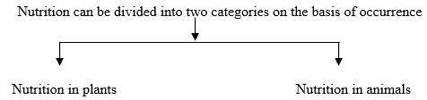
3. NUTRITION IN PLANTS
- Plants are autotrophic in nature. They prepare their own food hence they are called as producers.
- They contain a green pigment called chlorophyll which can entrap solar energy which is then converted into chemical energy in the form of food and the process is called as “Photosynthesis”.
Photosynthesis
(i) The synthesis of organic compounds like glucose from simple inorganic molecules like CO2 and H2O by the cells of green plants having chlorophyll in the presence of sunlight is called as photosynthesis.
(ii) Equitation of photosynthesis : Photosynthesis is a two step process.
(A) Light reaction : AT, NADPH2 and O2 are produced.
(B) Dark reaction : CO2 & H2O are converted into glucose.
Photosynthesis essentially requires two things :
Sunlight
- For plants sun the basis source of radiant energy.
- Plants utilize the light in the visible region of solar spectra (electromagnetic spectrum) which comes under the range of 390 nm – 780 nm.
- Visible region consists of white light which is a mixture of 7 lights of different wavelengths.
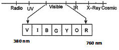
- Maximum photosynthesis occurs in red region.
- There is minimum photosynthesis in green region because green parts of plants reflect whole of the green light.
Chlorophyll
These are the green pigments present in chloroplast. They are found in green leaves in a maximum amount as well as in other green aerial parts of plant. There are six different types of chlorophyll, they are chlorophyll a,b,c,d,e and bacteriochlorophyll, amongst them chlorophyll a and chlorophyll b are the most commonly occurring chlorophylls.
Besides chlorophyll certain other pigments are also present in plants like.
(i) Carotenes : Orange in colour e.g. Carrot.
(ii) Xanthophylls : Orange yellow in colour e.g. Maize.
(iii) Phycobilins : Different colour like red, violet e.g. Blue-green algae, brown algae etc.
Raw Materials of Photosynthesis
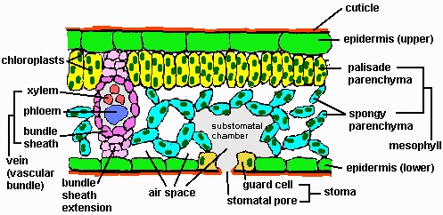
(i) Carbon dioxide : Terrestrial plants obtain carbon dioxide from the atmosphere through the small openings present on leaves called as stomata. ‘Stomata’ are the small pores present on the surface of leaves. They help in exchange of gases and water. Stomata opening is guarded by the presence of guard cells (kidney shaped). Aquatic plants obtain CO2 dissolved in water through their general body surface so they perform more photosynthesis than terrestrial plants. 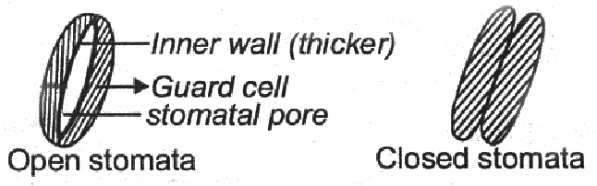
(ii) Water : Plants absorb water from the soil by the process of osmosis. This water is transported to leaves by a special type of tissue called as xylem.
- Plants utilize carbon dioxide during photosynthesis, the intensity of light at which amount of CO2 used during photosynthesis becomes equal to the amount of CO2 released during respiration by plants in called as Compensation point.
- Compensation point occurs at low light intensity that is during morning and during evening hours.
Site Photosynthesis Site of photosynthesis is different in prokaryotes and eukaryotes.
- In prokaryotes : Photosynthesis occurs in lamellar chromatophores.
- In eukaryotes : Photosynthesis occurs in chloroplast.
- Exception : Fungi ( It lacks chlorophyll so no photosynthesis occurs here).
- In higher plants chloroplast in the main site of photosynthesis.
- Chloroplast is also called as green plastid.
- Plastid was first observed by Haeckel.
- Plastids are of 3 different types on the basis of pigments present in them.
(i) Leucoplast: White in colour, found in underground parts, lacks and coloured pigment. Helps in storage of protein (Aleuroplst), oil (Elaioplast), starch (Amyloplst)
(ii) Chloroplast: Colour other than green found in aerial parts on the plants
(iii) Chloroplast: Contain green pigment, called as chlorophyll.
- Chloroplast was discovered by Schimper.
- Number of chloroplasts is variable in different species of plants.
- In lower plants like algae they are 1 or 2 number.
- In higher plants their number varies from 40 -100 per palisade cell or more.
- Chloroplast also have variable shapes, for example cup shaped, ribbon shaped etc. in algae while it is discoidal in higher plants.
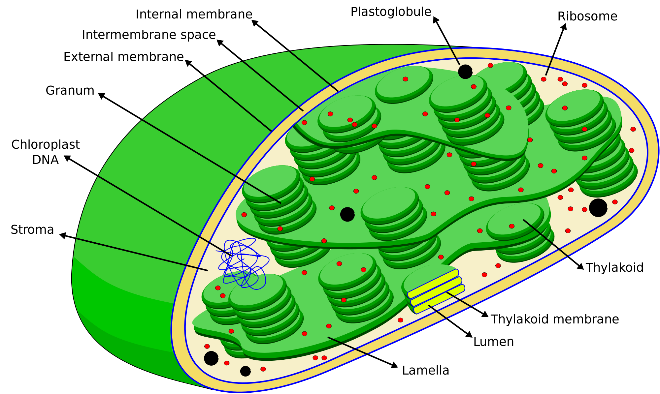
- A typical structure of chloroplast is a double membranous structure having two parts.
(i) Grana : It is a lamellar system consisting of stacks of granum lamella each bounded by a membranous box called as thylakoid. They are 40 – 60 per cell. Number of thylakoids per grana is 50 or more Chlorophyll molecules are found inside the thylakoid membrane where they trap solar energy in the form of small energy packets called ‘photon’ or ‘quanta’. Grana are interconnected to each other by a channel called as stroma lamellae or Fret’s channel.
(ii) Stroma : It is a non pigmented proteinaceous matrix in which grana remain embedded. It contain enzymes for dark reaction.
Mechanism of Photosynthesis
(i) Light reaction :
- It is also called as photochemical process.
- It was discovered by ‘Robert Hill’ therefore it is also called as Hill’s reaction
- Site : Grana of chloroplast.
- Raw materials : Light and water.
- Regulation : This process is regulated by chlorophyll molecules.
It consist of 3 steps :
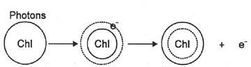
(A) Photo excitation of chlorophyll molecule : During this process chlorophyll molecule receives sunlight in the form of small energy bundles called as photons and become excited to higher energy level.
(B) Photolysis :
It is also called as photoxidation of water, this takes place in presence of Mn+2 and Cl– ions.
is liberated as by product and ions are used for the reduction of NADP
(C) Photophosphorylation: During this process ATP are produced. It takes place in quantasomes.
Mg+2 ions and inorganic phosphate is required to convert ADP ATP, ADP + 1P ATP.
(ii) Dark reaction :
- It is also called as thermo chemical reaction.
- It was discovered by Melvin calving and Benson therefore it is also called as Calving cycle Site = Stroma of chloroplast.
- Raw materials : They require CO2, , ATP, and Enzymes.
- Regulated by : Light reaction and enzymes.
- It involves three basic steps :
(A) Carboxylation : In this step CO2 is captured by CO2 acceptors like RUBP (C3 Plants) PET ( Plants) with the help of carboxylase enzyme i.e. RUBISCO & PEPCO respectively.
(B) Synthesis : This phase cap true CO2 is assimilated into glucose in the presence of phosphatase and isomerease enzymes and RUBP is regenerated back.
(C) Regeneration of RUBP
4. FACTORS AFFECTING PHOTOSYNTHESIS Light
Normally plants utilize sunlight but marine algae can perform photosynthesis even in the moon light. Plants can also perform photosynthesis in the artificial lights.
- Highest rate of photosynthesis : Red light
- Minimum photosynthesis : Green light
- Very high light intensity can cause reduction in the rate of photosynthesis by causing
(i) Decrease in transpiration rate
(ii) Denaturation of chlorophyll molecule
Temperature
Optimum range = 25° C to 30° C
It ranges from 10°- 40° C
In some forms like algae of hot spring 60° – 70° is normal
Carbon dioxide
It is the first limiting factor 0.03 – 0.1% is present in the atmosphere concentration of CO2 rate of photosynthesis.
above 0.9%
between 0.1 to 0.9%, it is constant and it is called as saturation point.
Oxygen
O2 acts as competitive inhibitor of CO2. Over concentration of O2 stops photosynthesis.
Chlorophyll
Chlorophyll content is directly proportional to rate of photosynthesis. No photosynthesis occurs in etiolated cells, In variegated leaves it occurs only at places where chlorophyll is present.
Significance of Photosynthesis
Photosynthesis is a boon to the nature and to the human beings. It has following significance :
(i) Production of food material(ii) Atmospheric control and purification of air.
5. NUTRITION IN ANIMALS
- Animals have highly evolved digestive mechanism that includes two basic components :
- Alimentary canal : Long, hollow, tubular structure consisting of various organs for digestion.
- Digestive glands : They secrete enzymes/hormones which help in digestion.
Digestion in animals consist of following steps :
- Ingestion : The process of intake of food.
- Digestion : It is the breakdown of large and complex molecules into simpler, smaller and soluble forms.
- Absorption : Taking up of the digested food through intestinal wall to blood.
- Assimilation : In this process absorbed food in taken by body cells.
- Egestion: The process by which undigested matter is expelled out.
- Digestive system is regulated by various hormones secreted by some endocrine glands.
- Alimentary canal was first of all developed in the phylum Platyhelminthes but only mouth was present in them.
- Coiled and well developed alimentary canal was developed in annelida till mammals.
Nutrition In Lower Animals
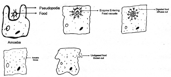
(a) Nutrition in Amoeba
It is a unicellular organism living in water.
- Mode of nutrition of holozoic.
- The process of obtaining food is the phagocytosis (cell eating)
- Steps involved in digestion of Amoeba are :
(i) Ingestion : Since it is unicellular so a single cell is responsible for carrying out all the vital activities. Food is ingested with the help of pseudopodia. Animal engulfs the food particle lying near it by forming pseudopodia around it and forming a food vacuole while is considered at its temporary stomach.
(ii) Digestion : The enzymes from surrounding cytoplasm enter the food vacuole and break down the food into smaller & soluble forms.
(iii) Absorption : The digested food is now absorbed by cytoplasm by simple diffusion and then the food vacuole disappear.
(iv) Assimilation : The food absorbed in Amoeba is used to obtain energy from respiration, for its growth and reproduction.
(v) Egestion : Undigested food is thrown out of the cell.
(b) Nutrition is Grasshopper
- It has a well developed digestive system having an alimentary canal and digestive glands.
- The various organs of digestive system of grasshopper are Mouth Oesophagus Crop Gizzard Stomach Ileum Colon Rectum.
- Glands associated with it are :
(i) Salivary glands
(ii) Hepatic caeca - Digestive system of a grasshopper can be divided into three parts.
(i) Foregut : mouth to gizzard
(ii) Midgut : gizzard to ileum (actual stomach)
(iii) Hindgut : stomach to anus.
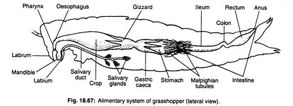
- The process involves:
(i) Ingestion : If feeds on green leaves so it takes food through its mouth with the help of it’s forelegs and mouth parts.
(ii) Digestion:
(a) It starts from the mouth.
(b) A pair of salivary glands secretes saliva and release it into the mouth through the salivary duct.
(c) Saliva mixed with food and lubricates and soften the food.
(d) Digestion of starch begins here.
(e) This slightly digested food enters into the crop through a food pipe i.e. esophagus.
(f) Crop stores the food temporarily.
(g) Now the food moves to gizzard. Here it is finally crushed and masticated an then moves to stomach.
(h) In stomach hepatic caeca release its secretions in the form of digestive enzymes, thus the food is then completely digested at this site.
(iii) Absorption : The digested food moves to small intestine (ileum) and absorbed through its walls.
(iv) Assimilation : Nutrients are assimilated whenever required by the cells for the fulfillment of the growth, energy and repair of the body.
(v) Egestion : Undigested food is then passed through hindgut (where H2O absorbed) and expelled out through anus in the form of elongated dry faecal pallets.
- The excretory organ of the grasshopper is malpighian tubules present at the junction of hindgut and midgut.
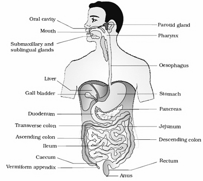
6. NUTRITION IN HUMANS
- Humans have highly evolved and complicated digestive system consisting of an alimentary canal and different types of digestive glands.
- Alimentary canal consist of following organs :
(a) Mouth :
It is small slit through which food is ingested.
(b) Buccal Cavity :
Mouth opens into a chamber called as buccal cavity. Roof of buccal cavity is called hard palate. At the floor of this cavity thick muscular structure is present called tongue. it helps in chewing swallowing, testing and speaking. Tongue has various types of papilla having taste buds.
- Jaws present in buccal cavity are provided with four different types of teeth :
(i) Incisors : For cutting
(ii) Canines : For tearing
(iii) Premolars : For grinding
(iv) Molars : For grinding
- Dental formula of humans :
(A) Milk teeth These are temporary, arise at 6 – 11 month age, 20 in number
(B) Permanent teetharise at 6 – 12 years, 32 in number
- Three pairs of salivary glands are found in mouth which release their secretions into the buccal cavity.
(c) Oesophagus :
Also called as food pipe. It leads the food from mouth to stomach, Oesophagus has highly muscular walls, no digestion occurs here.
(d) Stomach :
It is a ‘J’ shaped bag present on left side of abdomen. It contains several branched and tubular glands present on the inner surface of its wall, which secret gastric juice.
(e) Small Intestine :
It is a coiled and narrow tube having 3 regions : Duodenum , jejunum, ileum.
- On the inner wall of small intestine numerous finger like projections are found which are called as villi, they increase the surface area of absorption.
- Duodenum is proximal part of small intestine receives secretion from liver and pancreas.
(f) large Intestine :
Small intestine opens into large intestine from w here the undigested food material is passed to anus through rectum. It is divided into three parts:
(i) Caecum,
(ii) Colon
(iii) Rectum
(g) Digestive Glands :
(i) Salivary glands : 3 pairs of salivary glands are found in mouth cavity. It helps in chemical digestion. They secret at enzyme called salivary amylase or ptyalin. It helps in digestion of starch.
(ii) Gastric glands : Present in stomach. They secret hydrochloric acid, protein digesting enzymes and mucus.
(iii) Liver : It is the largest gland, secrets bile into the small intestine. Bile contains bile juice and bile pigments. Bile is alkaline in nature and it is temporarily stored in gall bladder and helps in digestion of fats, it also helps in absorption fats.
(iv) Pancreas: It lies parallel to and below the stomach. It secrets pancreatic juice into small intestine. Pancreatic juice contains tyrosine and pancreatic amylase. Besides these 2 enzymes pancreas secretes 2 hormones also i.e. :- insulin and glucagons so it has both exocrine as well as endocrine functions.Both bile and pancreatic juice are released into the duodenum by a common duct.
Intestinal Glands
They secrete intestinal juice and mucus.
Digestive System
This system involves following processes :
(i) Ingestion : Intake of food is done through mouth, food is then chewed and masticated and sent to esophagus through pharynx by swallowing.
(ii) Digestion : Saliva secreted in buccal cavity starts digestion of starch into maltose. This partly digested food is then passed to stomach by esophagus through peristaltic movement. Food is churned in stomach for about three hours and broken down into smaller pieces. Due to presence of hydrochloric acid, medium of stomach becomes acidic. In acidic medium protein digestive enzyme pepsin breaks down proteins into peptones. Gastric Lipase is also secreted here which partially breaks down lipids.
- Secretion of gastric juice is stimulated by the sight, smell or thought of food.
- Now the partly digested food moves to small intestine i.e. in the duodenum. Duodenum receives the secretion from liver and pancreas through a common duct they are bile and pancreatic juice, and alkaline in nature. So the digestion and emulsification of facts occurs at this place.
-
Here in the duodenum fats are emulsified by bile, remaining proteins are digested by trypsin and starch by pancreatic amylase.
NOTE : Duodenal wall secretes bicarbonate ions which make the medium alkaline.
- This partially digested food now enters in the ileum where intestinal juice i.e. “Succus entericus” is secreted. At this place digestion is completed.
Carbohydrates Glucose
Proteins Amino acids
Fats Fatty acids and glycerol
(iii) Absorption : After digestion molecules are broken down into simpler water soluble forms now they are to be utilized, so they pass through the wall of small intestine which contains blood capillaries and enters into the blood. For absorption of fat lymph capillaries are present called as lacteals.
NOTE : Wall of small intestine have tiny finger like projections called villi, they increase the surface area for absorption.
(iv) Assimilation : The process of utilizations of food is called assimilating. The nutrients dissolved in blood are carried to all parts of the body where they are utilized.
(a) For building up and replacement of cells.
(b) For obtaining energy. This energy is released by the process of oxidation during respiration.
(v) Egestion : The undigested food is then collected in large intestine where water is absorbed and remaining waste is expelled out or egested through anus.
RESPIRATION
1. RESPIRATION
The sum total of all the vital activities is called as metabolism. Vital act ivies refer to all the physiochemical activities of a cell. It has two aspects :
(i) Anabolism : It includes metabolic process by which complex cellular compounds are synthesized from simpler compounds, .e.g. Photosynthesis
(ii) Catabolism : It includes metabolic processes by which larger molecules are broken down into simpler molecules, e.g. Respiration. Respiration is an important catabolic process responsible for the production of energy.
Definition
The process by which assimilated food is oxidized and energy is released is called as respiration. In this process oxygen from air is taken in, this oxygen reacts with food molecules present in the body cells and burn them slowly to release energy. This energy is stored in the form of ATP molecules inside the cell for further use and the waste products i.e. CO2 and H2O are eliminated out of the body.
Food + oxygen CO2 + H2O + Energy
It is called as aerobic respiration.
NOTE : The process by which organisms obtain oxygen from environment and release carbon dioxide produced during oxidation of food to the outer environment is called as breathing. It is a part of respiration.
Difference Between Breathing and Respiration
(i) Breathing involves taking in of oxygen and releasing out of carbon dioxide so it is a physical process while respiration is a biochemical process which, along with breathing involves oxidation of food.
(ii) Breathing involves lungs so it is an organ system level process while respiration besides being at organ system level, also occurs at cellular level.
(iii) Breathing itself do not release energy while respiration results in the release of energy which is then stored in from of ATP.
(iv) Breathing is a part of respiration while respiration is not a part of breathing but it involves breathing.
Types of Respiration
(i) External respiration : Exchange of gases between an organism and its environment.
(ii) Internal respiration : Exchange of gases between tissue cells and extra cellular environment.
(iii) Aerobic : When oxidation of food takes place in presence of molecular oxygen.
it is called as aerobic respiration.
(iv) Anaerobic respiration : When oxidation of food material does not require molecular oxygen or it occurs in absence of molecular oxygen, it is called as anaerobic respiration.
Respiration
Respiration in divided in three parts :
(i) Cellular respiration
(ii) Respiration in plants
(iii) Respiration in animals
2. RESPIRATION PLANTS
-
- In plants exchange of gases takes place from leaves, stems and roots individually.
- Transfer of respiratory gases from one part to another is very less.
- Exchange of gases in plants occurs by simple diffusion.
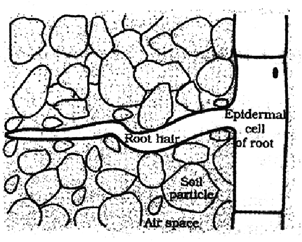
(i) Respiration in roots :
- In young roots, the epidermal cells are extended to form root hair. These root hair remain in direct in contact with the air present in between the soil particles. The oxygen from this air enters into the root hairs by simple diffusion and reaches to other cells of root for respiration.
- In older roots a protective layer of dead cells is present which have tiny openings called as lenticels. Diffusion of oxygen takes place through these pores and carbon dioxide is released out through the same.
(ii) Respiration in stem :
- In herbaceous plants, stem have small openings in their epidermal cells called as stomata, the oxygen from air enters through stomata and carbon dioxide is released from the same.
- In hard and woody stems of big plants and trees, lenticels are present in place of stomata through which exchange of gases takes place.
(iii) Respiration in leaves :
-
- Surface of leaves possess numerous tiny pores called as stomata in their epidermal cells, exchange of gases takes place through stomata and when CO2 concentration in cell increases stomata opens and CO2 is released out.
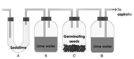
- An experiment to show that plants take oxygen and evolve carbon dioxide during respiration :
- Experiment : To demonstrate the plants take oxygen and evolve dioxide during respiration set the apparatus according to figure by taking KOH in U-tube, lie-water in two wide mouth bottles, one potted plant, bell jar and black-cloth. During day time the potted plant is covered with black-cloth to check photosynthesis. Make the apparatus airtight and start the aspirator. After sometime you will find that the lime water of second bottle turns milky. The explanation for this is that when the water comes out from aspirator, the atmospheric air enters into the apparatus through the second end and passes through the U-tube containing caustic potash into the tube containing lime water. The caustic potash absorbs the CO2 of the air. Thus, CO2 free air reaches into lime water so it does not turn milky. If indicates the air does not contain even trace of CO2. When this air reaches into the lime water of second tube through a bell jar having potted plant covered with black cloth to check photosynthesis, it turns milky. It proves that CO2 is evolved during respiration.
3. RESPRITAIN IN ANIMALS
-
- Respiration in animals takes place as a single unit, they have different types of organs for respiration due to which mode of respiration also varies according to the organism but the basic mechanism is same.
- From phylum Protozoa to Ctenophore respiration is by generally body surface, in phylum Platyhelminthes to Nematodes are mostly anaerobic and endoparasites, in phylum Annelida cutaneous membrane occurs and then from phylum Arthropoda till Mammals various respiratory organs were developed like trachea, gills and lungs.
|
Type of respiration |
Organs involved |
Example |
|
1. Cell surface respiration |
General body surface |
Amoeba, Paramecium |
|
2. Tracheal respiration |
Trachea and tracheoles |
Insects |
|
3. Branchial respiration |
Gills |
Fishes |
|
4. Cutaneous respiration |
Skin |
Frog |
|
5. Pulmonary respiration |
Lungs |
Amphibians, reptiles, birds |
|
6. Buccal respiration |
Buccal cavity |
From |
Some important characteristics of respiratory organs of animals are :
- They have large surface area to get enough oxygen.
- They have thin walls for easy diffusion and exchange of gases.
- They have rich blood supply for transport of respiratory gases.
Respiration in Amoeba
In unicellular organisms like Amoeba and in some lower multicellular animals likes sponges and cnidarians, respiration or exchange of gases occurs through general body surface as these cells are in direct contact with an aquatic environment so the oxygen dissolved in water diffuses into the cell and brings about oxidation of food, at the same time carbon dioxide released is expelled out of the cell by the same process.
Respiration in Earthworm
In organisms like earthworm and leech exchange of gases occur through their skin as their skin is very thin and most. It is rich in blood supply so the oxygen is absorbed by moist skin of earthworm and is transported to all the cells of body through blood. The carbon dioxide from body cells diffuses into the blood and expelled out through skin.
Respiration in Fish
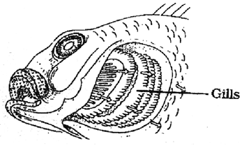
• In fish exchange of gases occurs through gills so the respiration is said to be branchial.
• Gills are present on both the sides of its head, they are covered by gill covers.
• During breathing fish takes in water through its mouth and pass it over the gills, the oxygen present in water extracted by gills and water is removed out through gill slits. This oxygen is now absorbed by blood and carried to all parts of the body and at the same time carbon dioxide is released into the blood and comes back to the gills and is expelled out into the surrounding water.
Respiration in Grasshopper
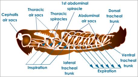
- In insects there occurs a system of tiny holes and air tubes all over the body these tiny holes or openings are called as spiracle. This whole system facilities the exchange of gases and is called as tracheal system.
- During breathing oxygen of air enters the spiracle and reached to each and every part of grasshopper’s body through trachea and tracheoles and carbon dioxide produced during respiration is carried back by trachea and tracheoles to the spiracles and is expelled out of the body of insect.
- The same mechanism is followed in other insects like houseflies, mosquitoes, bees etc.
4. RESPIRATION IN HUMANS
- Human respiratory tract
(i) External nostrils : First part of respiratory system. It opens into nasal cavity and is meant for inhalation of air from outside.
(ii) Nasal cavity : This cavity is separated from oral cavity by means of a hard and bony palate. It is lined by ciliated columnar epithelial cells that are rich in mucus; it brings about warming, moistening and sterilization of air. It contains hair and mucus which entrap the dust particles.
(iii) Internal nares : Nasal cavity opens into it and it leads to pharynx.
(iv) Pharynx : It is a common part between both alimentary canal and respiratory system.
(v) Larynx : It is an enlarged part of trachea which is also called as ‘voice box’. It produces voice by passage of air between vocal cords. It contains four different types of cartilages among them a ‘c’ shaped thyroid cartilage protruding out in neck region is called Adam’s apple.
(vi) Trachea : also called wind pipe. It is 10-12 cm long tube. It’s walls are supported by 16 – 20 ‘c’ shaped cartilaginous rings which percent them to collapse when air is absent in them
(vii) Bronchi : Trachea is branched into two bronchi left and right each of which enters into the lungs.
(viii) Lungs : These are two light weight spongy pouches covered by a membrane called Pleura. Bronchi are further branched into several bronchioles, at the end of bronchioles alveolar sacs or alveoli are present which are rich in blood capillaries and thin walled.
(ix) Diaphragm : It is a sheet of muscles that lies below the lungs and separates thoracic cavity from abdominal cavity.
- Mechanism of breathing : It includes
(i) Inhalation : When air is breathed in, the diaphragm and muscles attached to the ribs contract due to which there occurs expansion of chest cavity, it results increase in volume of chest cavity thus the air pressure decreases and air from outside rushes into the lungs and alveolar sacs get filled with air containing oxygen. The oxygen present in air diffuses into the blood and CO2 from blood diffuse out into alveolar sac.
(ii) Exchange between blood and tissues : CO2 is taken by blood and O2 diffuses into tissues.
(iii) Exhalation : When air is breathed out the diaphragm and muscles attached to ribs relax, which brings about contraction in chest cavity, its volume gets reduced and CO2 is pushed out from lungs into the air through trachea and nostrils.
Mechanism of gaseous exchange between tissues and blood
When the air enters into the lungs through nostrils, trachea and bronchi it enters into the bronchioles, from bronchioles it moves into thin walled alveolar sacs or alveoli. Alveoli are rich in blood capillaries, at this place oxygen from air diffuses into the blood and reaches to all the cells and tissues of body this oxygen now diffuses into the cell and is utilized for the oxidation of food and production of energy in mitochondria as a result of this carbon dioxide is produced in cells, due to this increased concentration of CO2, it diffuses into the blood and is brought back to alveoli and expelled out of the lungs through trachea and nostrils. 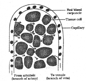
Control of Respiration
Respiration is controlled by the respiratory centre situated in medulla oblongata of brain.
(i) Breathing occurs involuntarily.
(ii) Under normal conditions rate of breathing is 15-18 times per minute. During vigorous exercise the demand for oxygen increases due to which rate of breathing increases by about 20-25 times.
(iii) The total area for gas exchange covered through 300 million alveoli is about 36-72 m2 in each lung.
(iv) Respiratory quotient : It is defined as the ratio of the volumes of CO2 liberated and O2 used during respiration.
5. SOME RESPIRATORY DISORDERS
- Emphysema : It occurs due to infection, smoking etc. It occurs due to obstructions in bronchioles caused by breaking of alveolar septa. Bronchodilators and O2 therapy are used, for curing this disease.
- Asthma : Air passages are narrowed and lead to obstruction in breathing.
- Pneumonia : Lymph and mucous accumulate in alveoli and bronchioles. It occurs due to bacterial and viral infection.
- Bronchitis : Swelling in living membranes of respiratory tract due to excessive smoking.
- Tuberculosis : Bacterial infection in lungs.
- Pleurisy : Inflammation of lung membrane called as pleurisy.
- Sudden contraction of diaphragm along with loud closure of glottis causes Hiccough.
- Sudden and violent expulsion of air through mouth and nose is called a sneezing.
- Fermentation : the slow decomposition of organic matter into simpler substances in the presence of enzymes is known as fermentation. This process is used for preparation of alcoholic beverages in presence of yeast in the absence of oxygen. Glucose and fructose are converted to ethanol by this process. It is a type of anaerobic respiration.
Difference Between Aerobic and Anaerobic Respiration
| Aerobic | Anaerobic |
| (i) It occurs in all living cells of higher plants. | It occurs in bacteria, certain fungi, germination seeds and fleshy fruits muscles. |
| (ii) It requires oxygen. | Oxygen in not required |
| (iii) The end products are CO2 and H2O. | The end products are alcohol or lactic acid and CO2. |
| (iv) The oxidation of one molecules of glucose produces 38 ATP molecules. | The number of ATP molecules produced is only 2. |
| (v) All the reactions except the reaction of glycolysis take place inside mitochondria. | All the reactions take place in cytoplasm. |
| (vi) Organic compounds are completely oxides and high amount of energy is released. | Organic compounds are incompletely oxidized and very small amount of energy is released |
| (vii) Non toxic to plants. | Toxic to higher plants. |
Differences Between Respiration and Photosynthesis
| Respiration | Photosynthesis |
| (i) It is a catabolic process. (ii) Carbohydrates are oxidized. (iii) Energy is liberated in the form of ATP. (iv) The amount of CO2 in the air increases during respiration. (v) It takes place in all the living cells, both green and non-green (vii) Dry weight of plant decreases. (viii) Oxidative phosphorylation occurs (ix) O2 is utilized and CO2 and H2O are formed C6H12O6 + 6O2 6O2 + 6H2O + energy |
It is an anabolic process. Light energy is stored in the form of glucose or chemical energy. It takes place only in chlorophyllous cells. |
Differences Between Respiration and Combustion
| Respiration | Combustion |
| (i) It is a biological process | It is a chemical process. |
| (ii) It takes place at normal temperature. | It takes place at high temperature. |
| (iii) Respiration is a slow process completed I n several steps. Thus, the energy is also liberated in several steps and remains stored in the form of ATP. | Combustion is fast process in which the energy is liberated only in one step resulting in increase in temperature and production of fire. |
6. CELLULAR RESPIRATION
If refers to the oxidation of food taking place inside the cell. As this process is at cellular level so it is called cellular respiration. It takes place in three steps :
It refers to the oxidation of food taking place inside the cell. As this process is at cellular level so it is called cellular respiration. It takes place in three in 3 steps :
(i) Glycolysis
(ii) Kreb’s Cycle
(iii) Electron Transport System
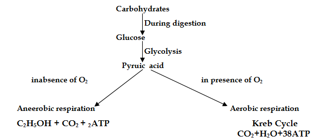
Glycolysis
Glycolysis also called EMP pathway, site-cytoplasm of cell.
(i) In this cycle glucose is converted into pyruvic acid in presence of many enzymes and co-enzymes.
(ii) Oxygen in not required during glycolysis.
(iii) 1 molecule of glucose gives rise to 2 molecules of pyruvic acid.
(iv) In this process 4 molecules of ATP are formed among them 2ATP molecules are utilized thus net gain of ATP is two molecules.
(v) 2NADP molecules are reduced to , which later produces 6ATP molecules.
(vi) Overall production of ATP in glycolysis is 2ATP + 6ATP = 8ATP
(vii) There is no production of CO2 during this process.
NOTE : After glycolysis, pyruvic acid is converted into acetyl Co-A with the release of CO2 and the process is called as ‘oxidative decarboxylation’. It occurs in mitochondria of the cell. Besides this 6ATP are also formed during this step.
Kreb’s Cycle
Site: Mitochondria of cell
(i) Also called aerobic oxidation.
(ii) Discovered by Sir Hans Kreb.
(iii) Another name TCA cycle (tricarboxylic acid cycle) or Citric acid cycle.
(iv) It brings about the conversion of pyruvic acid, fatty acids, fats and amino acids into CO2 and water by oxidation.
(v) It is the common path for oxidation of carbohydrates, fats, proteins.
(vi) it accounts for 24ATP molecules.
(vii) It starts with acetyl Co-A which is then converted into several intermediate compounds with the release of hydrogen atoms and then Acetyl Co-A is regenerated back.
(i) Electron Transport System or ETS
(i) In this hydrogen atoms produced during oxidation of various intermediates during Kreb cycle are first broken into protons and electrons.
(ii) These protons and electrons after passing through a series of coenzymes and cytochromes combine with oxygen to form water molecules.
(iii) During these series of events eleases 3ATP molecules and gives 2ATP molecules which were produces during Kreb cycle and glycolysis.
NOTE : The net gain of ATP molecules during respiration in 38ATP molecules among them,
8ATP from glycolysis
6ATP from conversion of pyruvic acid into acetyl CO.
A 24ATP from Kreb cycle
besides this CO2 and H2O are also released
Some important points
(i) Diaphragm becomes flat during inspiration and becomes convex during expiration.
(ii) Tidal volume : Volume of air inspired or expired in relaxed position. It is around 500 ml.
(iii) Residual volume : Air left in the whole reparatory tract after forceful expiration. It is 1.5 liters.
(iv) Total lung capacity : Maximum amount of air the lungs can hold after forceful inspiration. It is about 5-6.0 litres.
(v) Vital capacity : Maximum amount of air which can be breathed out through forceful expiration after a forceful inspiration. It is 3.4-4.8 litres.
- Vital Capacity is more in athletes, mountain dwellers, non smokers.
- The total area for gas exchange provided by our 750 million alveoli in two lungs in 100 S. m.
- In the cycle of inhalation and exhalation, repeated 15 to 18 times in a minutes about 500 ml of air is breathed in and out. In 24 hours, we breadth in 1500 litres of air.
- Blood is the medium for the transport of oxygen from the respiratory organ to the different tissues and carbon dioxide from tissues to the respiratory organs. As much as 97 percent of the oxygen is transported from the lungs to the tissues in combination with hemoglobin and only 2 percent is transported in dissolved condition by the plasma.
- A normal person has about 15 grams of hemoglobin per 100 ml of blood. One gram of hemoglobin binds about 1.34 ml of O2. Thus, 100 ml of blood carries about 20 ml of oxygen.
- Carbon dioxide is also transported by hemoglobin. When a respiring tissue release carbon-dioxide, it is first diffused in the plasma. From here it diffuses into the red blood cells. Carbon-dioxide is transported from the tissues to the lungs in the form of bicarbonates dissolved in water.
- About 23% of carbon dioxide entering into the erythrocytes combines with the globin (protein) part of haemogloin to form carbaminohaemoglobin, which is transported to the lungs.
- Carbon monoxide binds with hemoglobin about 230 times more readily than oxygen. When a person inhales carbon monoxide, it diffuses from the alveolar air to the blood and binds to haemoglobin forming carboxyhemoglobin. The latter is a relatively stable compound and cannot bind with oxygen molecules. So, the amount of hemoglobin available for oxygen transport is reduced. The resulting deficiency of oxygen cases headache, dizziness, nausea and even death.
- Mountain sickness : It is also known as altitude sickness. At sea level the concentration of oxygen is about 21% and the barometric pressure averages 760 mm Hg. As altitude increases, the concentration remains the same but the number of oxygen molecules per breath is reduced. AT 12,000 feet the barometric pressure is only 483 mm Hg, so there are roughly 40% fewer oxygen molecules per breath. In order to oxygenate the body effectively, breathing rate (even while at rest) has to be increased. This extra ventilation increases the oxygen content in the blood, but not sea level concentration. The fall in oxygenation of blood produced the symptoms of mountain sickness. These symptoms include breathlessness, headache, dizziness, nausea, vomiting, mental fatigue and a bluish tinge on the skin, nails and lips.
TRANSPORTATION
1. INTRODUCTION
All living bodies need nutrients and oxygen in every cell of its various tissues to sustain life. The transport of different material and gases is essential both in plants and animals. Unicellular organisms e.g. Amoeba and Paramecium do not require the transport of any material. These are in direct contact with their surroundings from where they obtain these nutrients. These substances are distributed in the cytoplasm due to the streaming movements of cytoplasm called as cyclosis. They exchange gases from the external environment directly by diffusion due to the difference in the concentration in and outside their body. In higher organisms both plants and animals, digested food, oxygen, hormones, waste nitrogenous substances etc. are to be carried from one place to the other. So transportation of materials is essential. It is done through circulatory system.
2. TRANSPORTATION IN HIGHER PLANTS
The higher plants have specialized system for the transportation of materials inside the body. The transportation of material is carried out by means of vascular tissues of the plants. The vascular tissues act as pipes or vessels. Through these vessels or pipes, water, minerals, salts, food etc. are transported in the plant body. In plants the medium of transportation is water. Water and food flows through the xylem (tracheids and vessels are the constituents of xylem) and phloem (sieve tubes and companion cells) for various metabolic activities. Tracheids and vessels are nonliving parts of xylem while sieve tubes and companion cells from the living parts of phloem. The terrestrial (land) plants absorb water and mineral salts through their roots. The area of young roots where most of the absorption takes place is the root hair zone. Root hair are the extensions of the epidermal cells. Root hair are delicate and do not live more than two days. The root hair have sticky walls by which they adhere tightly to soil particles. The root hair absorb water from soil by the process of osmosis but take in mineral salts by diffusion. The water and mineral salts are transported from the roots to the leaves, flowers and other parts of the plant. The upward movement of cell sap (water and minerals) through the xylem is called “ascent of sap”.
Translocation
Phloem translocates the manufactured food (sugar) or starch from the leaves to the leaves to the different parts of the plant including the roots.
Transpiration
Most of the water absorbed is lost through the aerial parts of the plant into air by a process called “transpiration”. Two percent of total water absorbed is used up in various metabolic activities in the plant body. Transpiration is the loss of water from the living tissues of the aerial parts of the plant in the form of water vapours. There are three types of transpiration :
(i) Cuticular transpiration (through cuticle)
(ii) Lenticular transpiration (through lenticels)
(iii) Stomatal transpiration (through stomata)
- Importance of transpiration :
(A) It controls the rate of absorption of water from the soil.
(B) It is responsible for ascent of sap.
(C) It regulates the temperature of the plant.
(D) Mostly water absorbed by roots is lost by transpiration without serving any purpose.
The energy spent by the plants in transpiration is wasted. So transpiration is a necessary evil.
Differences in Function of Xylem and Phloem :
|
Xylem |
Phloem |
|
(i) Functional xylem ells are dead. |
(i) Functional phloem cells are alive. |
|
(ii) It carries mineral salts, water and traces of organic molecules |
(ii) An organic solution of sugars and amino acids is translocated |
|
(iii) The movement is only upward. |
(iii) The movement can be upward or downward. |
3. TRANSPIRATION COHENSION THEORY
The main loss of water is through stomatal transpiration. Turgor pressure in the mesophyll cells of the leaf forces water outwards through the cell wall. Water evaporates from the surface of the cells into the air spaces of the spongy tissues and then passes into the outer atmosphere through the pores or stomata. The cell sap of mesophyll cells becomes concentrated by losing water and causes ad drop in turgor pressure. As a result water is sucked from adjoining mesophyll cells and ultimately from vascular tissues. This tension is transmitted all the way down to the unbroken column of water through the stem to the absorbing parts of the root. The molecules of the water show cohesion (mutual attraction) and molecules of water and vessel wall show adhesion (affinity for water). Due to these adhesive and cohesive forces, water column does not break but pulled upward by the force called as “transpiration pull”. The whole process can be compared with a person (transpiration pull) pulling a bucket full of water (forces on water column) from a well with a rope (column of water due to cohesion).
4. TRANSPORTAION IN HUMANS
In humans there is a circulatory system that uses blood or lymph as carries of materials (fluid exchange medium) and the heart as the pumping organ to help in circulation. Circulatory system consists of blood vascular system (blood as carrier) and lymphatic system (lymph as carrier).
Blood Vascular System
The higher multicellular animals with higher metabolic rates posses a well developed blood vascular system. This system helps in the quicker supply of nutrients and oxygen to the body tissues and also in the rapid disposal of toxics waste material and carbon dioxide. The blood acts as the circulatory fluid. Blood vascular system consist of blood, blood vessels and heart.
(i) Blood : The blood is a specialized kind of living connective tissue which is made to circulate, by the muscular pumping organ called as the heart. In adult human beings there is 5.5 to 6 liter of blood. The blood consists of fluid part, the plasma. The red blood corpuscles (RBSs), white blood corpuscles (WBCs) and blood platelets are present in the plasma. The formation of blood is called “Hempieces”.
(ii) Plasma : The plasma consist of water (90% & above) inorganic substances. In the plasma RBCs, WBCs and blood platelets float. Inorganic salts (09%) are also present. The organic substances are glucose, amino acids, proteins, hormones, digested and waste excretory products. The blood proteins (7%) are fibrinogen, albumin, globulin and prothrombin.
NOTE : Serum is plasma from which fibrinogen is removed.
(A) Red Blood Corpuscles (RBCs) or Erythrocytes : The number of RBCs is about 5.5 million in 1 ml of blood. The total number of RBC is about 30 billion. Each RBC is a biconcave disc-like structure devoid of nucleus. The mammalian erythrocytes do not possess nuclei, mitochondria and endoplasmic reticulum. The erythrocytes contain hemoglobin. Hemoglobin consist of globin (protein) and F2+ porphyrin compels (haeme). 100 ml of blood contains 15 mg of hemoglobin. if the amount of hemoglobin in blood is less, the person suffers from anemia The hemoglobin carries oxygen to the different cells of the body and brings carbon dioxide from the cells. The life span of a RBC is 120 days.
(B) White Blood Corpuscles (WBCs) or Leucocytes : The number of leucocytes is comparatively fever i.e. one ml of blood contains 5000 – 10000 leucocytes in humans. The total number of WBCs is about 75 millions. The number of leucocytes increases in infections like pneumonia, blood cancer (Leukemia) etc. These are large in size and contain nucleus. White blood corpuscles are of two types :
- Granulocytes : In granulocytes the cytoplasm contains granules and the nucleus is multilobed. Bosophils, Eosinophils and Neutrophils are three different types of granulocytes. Eosinophils and neutrophils are phagocytic (engulf and kill harmful microbes ) in nature and this process is called as “phagocytosis”. The function of basophils is to release histamine and Heparin.
- Agranulocytes : Monocytes and lymphocytes are two different types of agranulocytes. Lymphocytes secrete antibodies which destroy microbes. The monocytes are phagocytic in nature.
(C) Blood platelets : These are small and without nuclei. Their number various from 0.15 to 0.45 million in 1ml of blood. Their normal life span is one week. These help in blood clotting at the site of injury by liberating thromboplastin.
Functions of Blood
Blood performs the following functions :
- Transpiration of nutrients : The digested and absorbed nutrients like glucose, amino acids, fatty acids are first transported to the liver and then to all the tissues for their storage, oxidation and synthesis of new substance.
- Transportation of respiratory gases : The respiratory gases (oxygen, carbon-dioxide) are transported by the blood. Oxygen is transported from the respiratory surface (lung, skin and buccal cavity) to the tissues and carbon dioxide from the tissues is taken to the respiratory organ for its removal.
- Transportation of excretory products : Different wastes from the different parts of the body are collected by the blood and then taken to the organs (kidneys, lungs, skin and intestine) from where they are exerted.
- Transportation of hormones : Hormones are produced by endocrine glands. These hormones have target organs (p lace to act). These are carried by the plasma of blood and bring about the coordination in the working of the body.
- Maintenance of pH : the plasma proteins act as buffer system and maintains required pH of the body tissues.
- Regulation of body temperature : The blood flows in all the parts of body, so it equalizes the body temperature. It carries heat from one place to another place in the body.
- Transportation of metabolic intermediates : The blood carries metabolic intermediates from one tissue to another for further metabolism. In the muscle cells due to anaerobic respiration lactic acid is produced. This lactic acid is carried to the liver for further oxidation.
- Water balance : The blood maintains water balance to constant level by distributing it uniformly in the body.
- Protection from diseases : The WBCs (eosinophils, neutrophils, monocyts) engulf the bacteria and other disease causing organisms by phagocytosis. The lymphocytes produce antibodies to neutralize the action of toxins produced by pathogens.
- Clotting of blood : Blood forms a clot at the site of injury and thus prevents the further loss of blood.
- Support : Blood flows under pressure in arteries. Due to this tissues become stiff as in the case of erection of nipples, clitoris and penis.
Blood Clotting
At the site of injury of the blood vessels, the platelets induce blood coagulation through the release of thromboplastin (thrombokinase). Thromboplastin changes prothrombin of blood plasma into thrombin. Thrombin converts soluble protein fibrinogen to insoluble fibrin. Fibrin forms a network which entangles RBCs and blood platelets to form plug or clot over the inured area. Blood clotting is usually completed within 2-3 minutes.
Blood Groups
Land Steiner discovered that blood of different individual did not match each other but there were biochemical differences. He discovered Antigens A and B and blood groups (ABO systems). Antigen (agglutinogen) is a glycoprotein present on RBCs. For each antigen there is a corresponding antibody. Thus there are two antibodies (agglutinin) a and b occurring in the blood plasma. There are four types of blood groups depending on the presence or absence of these antigens.
|
Blood Group : Antigen and Antibody |
||
|
Body Group |
Antigen present on RBCs |
Antibody in plasma |
|
A |
A |
b |
|
B |
B |
a |
|
AB |
AB |
None |
|
O |
None |
a,b, |
Blood is a life saving fluid. It is often needed during accident and operation. The transfusion of blood is only done when blood group is known. These groups are A,B,AB and O. Blood of O group is a universal donor i.e. donate blood to any group (a, AB, B and O) but it can receive blood from O blood group. A B group is universal recipient (receiver). It can receive blood from any group (A, B, AB, O) but it can donate to AB group only.
Blood Transfusion
The transfusion of blood from a healthy person to a patient suffering from blood loss due to injury or surgical operation is called a “blood transfusion”. For this all major hospitals have blood banks where blood is collected from voluntary and professional donors. Before preservation the blood is tested for its blood group and Rh factor. Though theoretically a patient may be able to receive blood of two or more types, it is always advisable to have the donor blood of the same group as that of the recipient. Rather the blood of donor is always cross-matched before transfusion to exclude any change of incompatibility. When blood from a donor is added is added to blood of the recipient, it is necessary to avoid bringing together corresponding antigen and antibody. This causes clumping of RBCs. Thus antigen A in RBCs of group A individuals reacts with antibodies of plasma of group B individuals. This phenomenon is called “agglutination”.
Table Human blood groups and transfusion
|
Blood group of donor |
Blood group of recipient |
|||
|
O |
O |
A |
B |
AB |
|
✓ |
✓ |
✓ |
✓ |
|
|
A |
✗ |
✓ |
✗ |
✓ |
|
B |
✗ |
✗ |
✓ |
✓ |
|
AB |
✗ |
✗ |
✗ |
✓ |
✓ Compatible
✗ Incompatible
Rh factor (in blood) can be genetically determined. Most of the people (more than 85%) are Rh positive () while a few are Rh negative (). Both people lead normal life. If an Rh- woman marries with an man then Its pregnancy is normal but in 2nd pregnancy the mother with blood may lose the baby due to incompatibility of Rh factor. By new techniques and procedures now the child can be saved.
5. STRUCTURE OF HEART
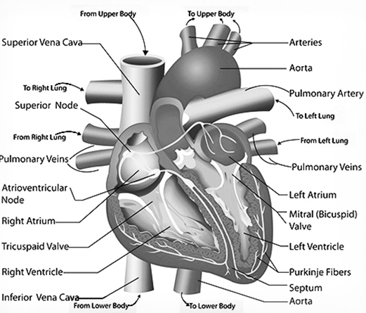
- Heart is a hollow muscular organ that lies obliquely in the thoracic region in a cavity between the two lungs that is pericardial cavity. It is lined by 2 layers outer and inner pericardial membranes. These are filled with a fluid called “pericardial fluid”. It protects the heart from shock and injury.
- Heart is made up of 4 chambers : upper 2 chambers are auricles and the lower 2 chambers are ventricles. Auricles are the receiving chambers and ventricles are the pumping chambers. Walls of ventricles are thicker as they have to pump the blood.
- Partition between right and left auricle is called “interauricular septum” and between right and left ventricles is “inter ventricular septum”.
- Four pulmonary veins enter into left auricle, two from each lung bring oxygenated blood. There is one auriculoventricular aperture with a bicuspid or mitral valve in left auricles which opens into left ventricle.
- Left ventricle has aortic valve having 3 semilunar cusps for large artery i.e. dorsal aorta which takes the oxygenated blood to all body parts.
- Right auricle has openings for superior venacava that brings deoxygenated blood from head, neck and upper limbs, inferior venacava receives deoxygenated blood from rest of the body and lower limbs. Blood enters into right ventricle through tricuspid valve. A coronary sinus that drains venous blood from heart muscles.
- Right ventricle has pulmonary valve having 3 semilunar cusps for pulmonary artery carrying deoxygenated blood to lungs.
- The series of events which occur during one heart beat is called as cardiac cycle.
- NOTE : During foetal condition a flap valve called “foramen ovale” is present at interauricular septum having a depression called as fossa ovalis. If it remains after birth it results “a hole in the heart”.
Blood Pressure
It is the pressure of the flow of blood in the aorta and its main arteries. The blood pressure varies according to the contraction and relaxation of the heart. In the condition of contraction or systolic phase (Lubb sound) it is about 120 mm of Hg. This is called “systolic pressure”. In the relaxation or diastolic phase (Dub sound) it is about 80 mm of Hg and is called “diastolic pressure”. The normal blood pressure of man (20 years) is 120/80. Fats and anxiety increase the blood pressure, the maximum normal blood pressure should into exceed 150 in males and 140 in females. The blood pressure is measured by “sphygmomanometer”.
Detection of Normalcy of Heart Beat
The muscle fibres of heart are specialized at certain parts generate tiny electrical currents which cause the normal heart heats. The “electrocardiograph” (E.C.G.) is the device to record these electrical changes. Electrocardiogram is a record of electrical behaviour of heart and remains constant in a normal man. Doctors use the E.C.G. for detection of various heart diseases. Sometimes the sinoatrial node (SA node or pacemaker) gets damaged and fails to generate cardiac impulses at normal rate it becomes abnormally slow and irregular and ventricles fail to pump the required amount of blood. It can be corrected by the surgical grafting of an artificial pacemaker instrument in the chest of the patient. This instrument stimulates the heart electrically at regular intervals to maintain the beats.
6. LYMPHATIC SYSTEM
The lymphatic system comprises the lymph, lymphatic capillaries (simply lymphatic), lymphatic vessels and nodes. Lymph severs as the middle man between the blood and organ for exchange of any material. The lymph is the tissue fluid present in the intercellular spaces in the tissues. So it is also called as “extracellular fluid”. The lymph resembles the blood except that the lymph is devoid of R.B.Cs, blood platelets and some plasma proteins. Lymphatic system runs parallel to the veins. The lymphatic capillaries are present in the form of network under epithelial surface. The ends of lymphatic capillaries are blind. The lymphatic capillaries unite to form lymphatic vessels and these vessels resemble with the veins. The lymphatic vessels posses the valves which prevent back flow of lymph. Neighboring body muscles help in the flow of lymph. The small lymphatic vessels unite to form large vessels. Larger lymphatic vessels unite to form large ducts i.e. right lymphatic duct and thoracic duct. Right lymphatic duct opens into right subclavian vein and left thoracic duct open in to left subclavian vein. Before the lymph reaches the blood, it always passes through the lymph nodes. The lymph’s nodes are enlargements of the lymphatic vessels. Lymphocytes and other plasma cells are present in the lymph nodes. The lymph is cleaned or filtered by lymph nodes. These cells also kill the germs and produce antibodies.
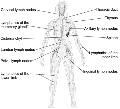
Functions of Lymph
(i) It provides immunity through lymphocytes.
(ii) Fats are absorbed through lymph vessels in the intestine
(iii) It supplies digested food and oxygen to various parts of the body.
(iv) It helps in removal of waste products like parts of dead cells.
(v) It returns proteins and excess tissue fluid to the blood from the tissue spaces.
EXCRETION
1. EXCRETION
There are various metabolic activities which take place inside the living organisms. All these activities are chemical reactions. As a result in animal body several end products are formed which are of no use to the cells. These are called as wastes. These must be removed from the body for proper functioning of the body. The elimination of these waste nitrogenous products from the body is called as excretion. Waste material is ammonia, urea, uric acid, carbon dioxide, pigments, salts digestive wastes, excess of water etc. Ammonia, urea uric acid are waste nitrogenous products, The excretory produces are both volatile and non-volatile. These are removed from the body by different methods.
Excretion in Amoeba
Amoeba is an ammonotelic organism since the principal excretory product is ammonia. Special excretory organelle in Amoeba is lacking. CO2 and ammonia are exerted by diffusing is solution through plasma membrane. The concentration of ammonia is always higher in Amoeba than in the surrounding water. The water enters through plasma membrane by “endosmosis”. Ammonia is formed in cytoplasm by metabolism. Surplus water enters contractile vacuole. This surplus water can rupture the animal’s body. Thus size of contractile vacuole increases, when the contractile vacuole is fully expanded with water, it moves towards the periphery. As it comes in close contact with the plasma membrane, the contractile vacuole bursts. Thus excess of water (surplus water) is discharged in the surrounding water, this phenomenon of controlling the amount of water in the body is called as “osmoregulation”.
Excretion in Earthworm
In earthworm, the excretory organs are nephridia. The internal funnel-like opening is called as “nephrostome”. The waste material from body cavity (coelom) enters the nephridium through nephrosome. In the inner lining of nephridium, the cells absorb useful substances like glucose.
Structure of a Typical Nephiridium
A typical nephridium consists of three parts : nephrostome, body and terminal duct. The nephridium communicates with the coelom (body cavity) through internal nephrostome. Nephrostome is a ciliated funnel which leads into body of nephridium through the neck. The body of nephridium consists of short straight lobe, a long spiral lobe with narrow apical part. Spiral lobe consists of proximal limb and distal limb. Neck of the nephridium leads into proximal part of spiral lobe and terminal duct leaves the proximal limb. The tubule of the neck enters the body of the nephridium and leaves the body as terminal duct. These tubules have ciliated tracts inside. The number of ciliated tracts depends upon the number of coils of the tubules. The terminal duct may open outside by nephridiopore or into the gut (alimentary canal).
Functioning of Nephridium
Nephridia are highly vascular and extract nitrogenous wastes from the blood. The nitrogenous wastes and useful substances (glucose) enter the body of nephridium through internal neprostome in the fluid form. The cilia present in the tubule beat to move the fluid. Useful substances like glucose are reabsorbed by cells, lining the tubule and is passed into the blood. The remaining waste is discharged into the alimentary canal or to exterior through nephridiopore. According to the position of nephridia in the body of earthworm, nephridia are of three types :
(i) Septal nephridia are attached on septa. Nephridiopore is missing.
(ii) Integumentary nephridia are attached on inner side of the skin. Nephridiopore is present.
(iii) Pharyngeal nephridia are present as three p airs of groups of nephridia, on both sides of alimentary canal. Nephridiopore is absent. Septal and pharyngeal nephridia are endonephric as these open in the alimentary canal. Integumentary nephridia are ectonephric. Excretion is an adaptation to conserve water. Earthworm is ammonotelic (excrete ammonia) in excretion, in sufficient water while it is ureotelic (excrete urea) on land.
2. HUMAN EXCRETORY SYSTEM
As a result of various metabolic process going on in our body a number of waste products are formed. These have to be eliminated as they are toxic to the body.
- The waste products include :
(i) Carbon dioxide which is liberated during respiration; and is eliminated by the lungs.
(ii) Nitrogenous metabolic wastes, such as urea and uric acid produced in the liver from excessive proteins.
(iii) Bile pigments : Bile pigments (e.g., Bilirubin) derived by the breaking down of hemoglobin of the erythrocyte.
(iv) Excess salts, water and vitamins : Concentration of these substance above the required level, is harmful to the body. Elimination of all metabolic nitrogenous wastes from the body is callers as excretion.
3. ORGANS OF EXCRETION
(i) Lungs : Carbon dioxide produced by the oxidation of glucose or other food substances in the tissues is removed by the blood. This carbon dioxide is carried to the lungs through the blood vessels (veins) where it diffuses into the alveoli and out through the respiratory tract. Water vapour in small amount is also exhaled during expiration from the lungs.
(ii) Skin : Substances like soluble food mater, oxygen, water dissolved mineral salts, traces of urea and uric acid diffuse from the thin walls of capillaries into the walls of the sweat glands. Oxygen and food substances are used for metabolic activities of the cells of sweat glands but the remaining metabolic wastes are excreted out of the gland through the sweat duct which opens on the surface of the skin through sweat pore. Sweat contains 99% water, traces of urea and uric acid. However, after heavy exercise, lactic acid forms a major constituent of seat. Profuse sweating may lead to sodium deficiency, leading to muscle cramps. An adaptation of prevention of water loss is the impermeability of our skin to water. However, in aquatic animals, skin is the major excretory organ. They excrete ammonia through their skin by diffusion as ammonia is highly soluble in water.
4. INTERNAL STRUCTURE OF KIDNEY
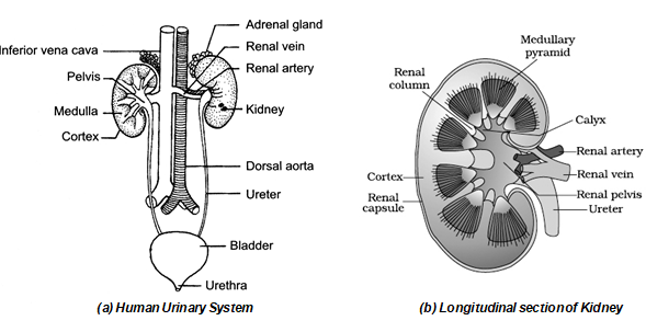
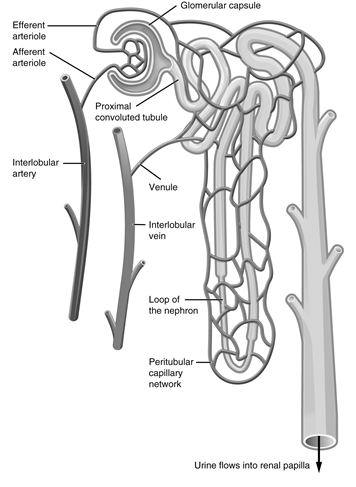
(i) Bowman’s capsule : It is a single-cells thick, double walled cup-shaped structure present in the cortex region of the kidney. The cup-shaped capsule contains a network of capillaries called Glomerulus’s. Glomerulus’s and Bowman’s capsule are together called as Renal corpuscle.
(ii) Proximal convoluted tubule (PCT) : It starts after the Bowman’s capsule and is greatly twisted. The
whole P CT lies in the cortex region.
(iii) Henle’s loop : Henle’s loops is a U-shaped tubule located in the medulla region. it consists of
(A) a thin-walled descending limb in the medulla
(B) a thick-walled ascending limb in the cortex. Henle’s loop is long in those animals which pass hypertonic urine.
(iv) Distal convoluted tubule : The ascending limb continues into the distal convoluted tubule which forms several coils in the cortex.
(v) Collecting duct : Collecting tubule receives distal tubules of several uriniferous tubules. Several such tubules unite to form a large collecting duct. The collecting ducts are held together and converge to form a pyramid. The pyramid opens into the pelvis which leads into the Ureter.
5. BLOOD SUPPLY TO NEPHTRONS
Inside the kidney, the renal artery branches into a number of renal arterioles. A branch from a renal arteriole enters each Bowman’s capsule, and is called the afferent arteriole. It breaks up into a network of capillaries which reunite to form a efferent arteriole. (Glomerulus is a mass of network of capillaries in the Bowman’s capsule). The efferent arteriole after emerging from the Bowman’s’ capsule runs a short distance and breaks up into a capillary network which surrounds the renal tubule and rejoins to form a vein. By reuniting again and again with other veins of the kidney it forms the renal vein which drains into the posterior venacava.
6. CHEMICAL COMPOSITON OF URINE
Normal human urine consist of about 95% water and 5% of solid wastes. Besides the normal constituents , certain hormones and medicines like the antibiotic and excess vitamins are passed out with urine. Organic compounds (gm/l): Urea – 2.3; Creatinine – 1.5; Uric acid – 0.7; Ammonia – 0.6 Inorganic Compounds (gm/l) Nacl – 19.0; KI – 12.5; H2SO4 – 1.8; NH3 – 0.6. Normally a man excretes 1000 – 170 ml of urine daily, depending upon the water intake, diet, climate, mental state and physiological condition. Tea, coffee, alcohol and other beverages increases the formation of urine.
Working of Nephron
Main function of nephron is to form urine. There are three main process involved in the urine formation :
(i) Glomerular ultrafiltration : If is the filtration of body fluids and solutes from the blood, out of the glomerular capillaries into the Bowman’s capsule due to the pressure in the glomerulus. All substances from the blood are filtered out except the large protein molecules. This fluid in the glomerular capsule is called as glomerular filtrate. It consists of water, urea, salts, glucose and other plasma solutes. Blood coming out of the efferent arteriole is therefore thick.
(ii) Tubular reabsorption : Glomerular filtrate contains a lot of useful materials like glucose, salts such as that of sodium and water. These substances are reabsorbed from the renal tubule at various levels and in varies proportions. Glucose is reabsorbed completely from the proximal convoluted tubule. More than 85% of water is reabsorbed from the proximal, distal and even in collecting tubules. Sodium chloride is reabsorbed in the proximal and distal tubules. Potassium and phosphate is completely reabsorbed from the proximal tubule. Other substances reabsorbed are uric acid, sulphates, vitamin C, amino acids etc.
(iii) Tubular secretion : This occurs mainly in the distal convoluted tubule and the collecting duct of the nephron. It is an active, vital process performed by the cells of the cuboidal epithelium lining the tubules which excrete additional wastes from the blood stream into the filtrate by active transport. In this process substances like potassium, hydrogen, creatinine and certain drugs like phenol, penciling etc. are directly exerted by the tubular cells from the blood. The fluid which now flows through the last parts of the tubule is urine which consist of water, urea, uric acid, mineral ions like sodium, potassium, chlorides, phosphates etc.
7. ARTIFICIAL KIDNEY
In case of loss or damage of one kidney, the other kidney performs the function of both the kidneys and the person can lead a normal life. But the failure of both the kidneys leads to death. Artificial kidney is a dialysis machine which cleans blood of waste products, thus acting like a kidney. The patients’ blood is led from the radial artery of the arm through the machine where urea and other salts are removed and pure blood is returned to vein in the same arm. In case of permanent damage to the kidneys, dialysis has to be performed for about twelve hours, twice a week. Patients with chronic kidney failure have been recorded to survive for more than 12 years on dialysis. Now a days, diseased kidney may be replaced with healthy one by kidney transplantation. To lead a normal life, one healthy kidney is more than enough. Therefore, a healthy person can donate his one kidney to patient who has both kidneys impaired.