


1. INTRODUCTION
- Food is the basic requirement of all living organisms as it provides energy and organic materials for growth and repair of tissues.
- Major components of food – carbohydrates, proteins and fats.
- Minor components of food – vitamins and minerals.
- Water plays an important role in metabolic processes and prevents dehydration of the body.
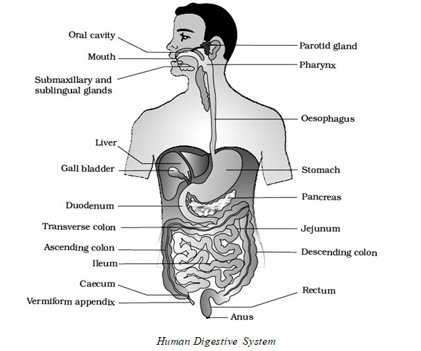
2. HUMAN DIGESTIVE SYSTEM
- Chemically it is a process of hydrolysis.
- The process of conversion of complex food substances to simple absorbable forms is called digestion and is carried out by our digestive system by mechanical and biochemical methods. It is of two types:-
- Intracellular digestion: Digestion within the cells e.g. Protozoans (Amoeba), Sponges, Cnidarians
- Extracellular digestion: Digestion outside the cells e.g. Cnidaria to chordata
- It consists of digestive tract or alimentary canal and associated glands.
- Total length of alimentary canal in man is 30 feet (6-9 meters) long.
Alimentary canal
- Begins with mouth and opens out through anus.
- Mouth leads into the buccal cavity or oral cavity. It is followed by pharynx, oesophagus, stomach, small intestine and large intestine.
Mouth and Buccal (Oral) cavity
- It is lined by non-keratinised stratified squamous
- Roof of buccal cavity is palate consisting of anterior hard palate posterior soft palate.
- Hard palate is made up of premaxilla, maxilla and palatine
- Thick transverse folds of mucus epithelium that lines the hard palate are called palatine rugae.
- Terminal part of soft palate that hangs in to the throat is called uvula.
- Buccal cavity has a number of teeth and a muscular tongue.
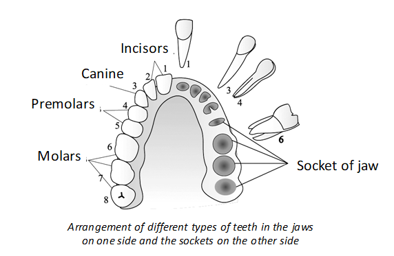
Teeth
- In human beings the dentition is thecodont, heterodont and diphyodont.
- Teeth are derived both from ectoderm and mesoderm.
- There are four types of teeth – incisors ( I ) for cutting, canines (C) for tearing, premolars (PM) for crushing, grinding and chewing and molars(M)for chewing, hence called heterodont.
- Except Premolars and last molars all the type of teeth appear twice in life hence called diphyodont.
- Teeth which appear during child hood are called milk teeth or primary teeth or deciduous teeth.
- Deciduous teeth consist of 20 teeth.
- All the deciduous teeth are lost generally between 6 to 12 years and are replaced by permanent or secondary teeth.
- The third molars are called wisdom teeth they appear only once at the age between 17 to 25 years.
- Canines and wisdom teeth are vestigial in man.
- There are no premolar teeth in milk dentition.
- Dental formula in human adult is , while milk dentition is
- The dental formula of monophyodont teeth is 0021 0021 i.e 0 Incisors, 0 Canines, 2 Premolars and 1 molar
- The maximum number of teeth in placental mammals with heterodont dentition is 44, e.g., horse and pig.
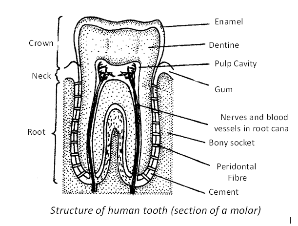
- Teeth are made mainly of dentine while the chewing surface of the teeth that helps in mastication of food is covered by enamel.
- Enamel is the hardest substance of the body. Enamel is made of calcium carbonate and calcium phosphate.
- It is secreted by ameloblasts of ectodermal
- Dentine is harder than bone and is secreted by odontoblasts which line the pulp cavity.
- Teeth are embedded in sockets of the alveolar processes of jaw bone, hence called thecodont.
- The alveolar processes are covered by the gingivae, or gums.
- The sockets are lined by the periodontal ligament or periodontal membrane made up of dense fibrous connective tissue.
- Tooth has three parts namely crown, root and neck.
- The crown is the visible part, neck is middle part and root is embedded in the socket of jaw bone.
- Tooth is mostly made up of bone like substance called dentine.
- Dentin of the root is covered by cementum.
- Cavity enclosed by root is pulp cavity filled with pulp.
- Narrow extensions of the pulp cavity are called root canals.
- Premolars and molars help in grinding and are called cheek teeth or grinding teeth.
- Thecodont : Teeth present in sockets.
- Diphyodont : Formed twice in the life time e.g., Incisors, canines and first two molars.
- Heterodont: Teeth of four different types such as incisors, canines, premolars and molars, characteristic of therian mammals.
Tongue
- Tongue is freely movable, muscular organ attached to the floor of the oral cavity by lingual frenulum.
- Tongue has striated extrinsic and intrinsic muscles.
- Tongue acts as universal tooth brush.
- The anterior two thirds are covered by lingual papillae.
- Three types of papillae are found on human tongue –
- Circumvallate (the largest, knob like and lie at the back of the tongue),
- Fungi form (mushroom shaped, Scattered over the entire surface of the tongue, contain tactile receptors but no taste buds.),
- Filiform (filament shaped, lie on the entire surface of the tongue.)
Pharynx
- It is a short passage for food and air.
- Structures that open into the pharynx are oesophagus and trachea (wind pipe).
- During the swallowing, entry of food into the wind pipe is prevented by epiglottis a cartilaginous flap.
- Pharynx is divided into three parts namely Nasopharynx (upper part), Oropharynx (middle part) and Laryngopharynx (lower part).
- Nasopharynx has internal nares, opening of the Eustachian tubes and a mass of lymphoid tissue called pharyngeal tonsil (adenoid).
- Oropharynx has an ovoid masses of lymphoid tissue called palatine tonsils.
- A pair of mass of lymphoid tissue called lingual tonsils are present at the base of tongue
- Pharynx leads into oesophagus.
Oesophagus
- The part of alimentary canal which passes through neck, thorax and diaphragm is oesophagus.
- It pierces through the diaphragm. It is ten inches long (25 cm) .
- It has both unstriped and striped muscles but lacks serosa.
- Opening of oesophagus into the stomach is regulated by gastro – oesophageal sphincter, also known as cardiac sphincter.
- Oesophagus opens into the stomach in the abdominal cavity.
Stomach
- The J-shaped stomach is located in the upper left portion of the abdominal cavity.
- It has four main parts cardiac, fundic, main body and pyloric portions.
- The wall of the stomach has three layers of muscles the outermost longitudinal layer, middle circular layer and innermost oblique layer.
- Mucosa of stomach forms large irregular folds, called rugae.
- Stomach leads into small intestine.
- Opening of stomach into duodenum is guarded by pyloric sphincter.
Small intestine
- Small intestine has three parts duodenum, jejunum and ileum.
- Duodenum is twelve inches (25cm) long and is ‘U’ or ‘C’ – shaped and receives hepatopancreatic duct.
- Pancreas lies between the two limbs of duodenum.
- Jejunum is 5 feet long and is a coiled part.
- Ileum is highly coiled and 5 feet long it is the longest and highly coiled region of the small intestine.
- The wall of ileum has Payer’s patches which produce lymphocytes.
- Small intestine leads into large intestine.
- Submucosa of the duodenum contains Brunners glands which secrete mucus.
Large intestine
- Large intestine consists of caecum, colon and rectum.
- It is 5 feet long and 2.5 inches in diameter.
- Caecum is a small blind sac, which has some symbiotic micro organisms.
- A vestigial organ arises from the caecum called vermiform appendix – three inches in length.
- Caecum opens into the colon.
- Colon is 5 feet and is divided into three parts as ascending, transverse and descending part.
- Descending part of colon passes into the rectum that opens into the anal canal.
- Anal canal opens the outside by anus which is guarded by external anal sphincter (made up of skeletal muscle) and internal anal sphincter (made up of smooth muscles).
- In certain conditions rectal veins can get distended or enlarged due to weakening of their valves leading to haemorrhoids.
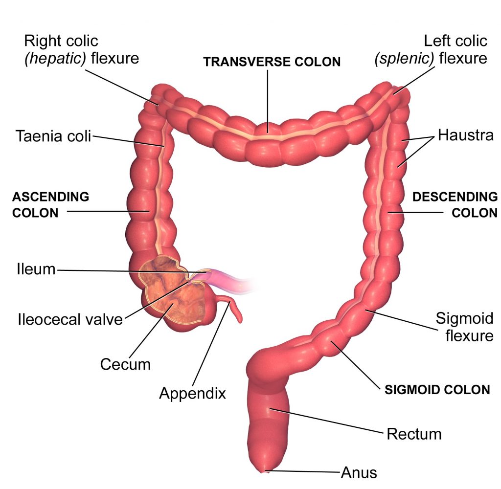
The Large Intestine
Histology of Alimentary canal
- Wall of the alimentary canal has 4 layers. Serosa 2.Muscularis 3.Submucosa 4.Mucosa.
- Serosa – Outer most layer and is made up of mesothelium and some connective tissue.
- Muscularis – Smooth muscles consisting of outer longitudinal and circular muscles. In some regions (stomach) oblique muscles are also present.
- Submucosa – Loose connective tissue and contains nerves, blood vessels and lymph vessels.
- In duodenum submucosa has ‘Brunner’s’ glands.
- Mucosa – It is the inner lining layer of alimentary canal. It has 3 parts the outer muscular mucosa (made up of longitudinal and circular muscles), middle lamina propria (made up of connective tissue, blood vessels, glands and lymphoid tissue) and inner most epithelium.
- It forms ‘rugae’ (irregular folds) in stomach.
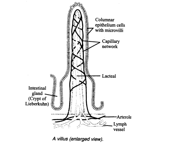
- It also forms villi which are small finger like foldings in small intestine.
- Cells lining villi bear microvilli which is seen as a ‘brush border’.
- Microvilli increase surface area of absorption enormously.
- Villi have blood capillaries and lymph capillaries called lacteals.
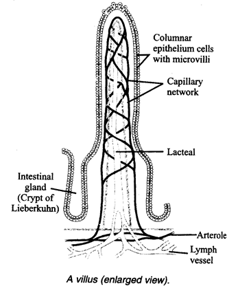
- Goblet cells of mucosa secrete mucus for lubrication during food passage.
- Digestive glands of stomach and crypts of Lieberkuhn of intestine are formed by mucosa.
3. DIGESTIVE GLANDS
- The digestive glands associated with alimentary canal include salivary glands, liver and pancreas.
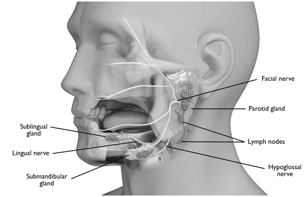
Salivary glands
- Human beings have three pairs of salivary glands.
1. Parotid 2. Submaxillary or submandibular 3. Sub lingual glands. - Saliva contains enzyme ptyalin.
- Ptyalin acts on starch and converts it to maltose in the presence of chloride ions.
- Serous cells secrete salivary amylase and mucus cells secrete mucus.
- Daily secretion of saliva is 1.5 liters. (pH is 6.8).
- Saliva has salivary amylase (alpha amylase or ptyalin), lysozyme, water and solutes such as Na+ K+ , and .
- ‘Mumps” is a viral disease caused by Paramyxo virus causing inflammation of parotid glands.
Liver
- It is the largest gland of the body.
- It weighs 1.2 to 1.5 kg in an adult human.
- It is located below the diaphragm in the abdomen.
- It has two lobes – right and left lobes.
- Structural and functional units of liver are hepatic lobules.
- Hepatic cells / hepatocytes are arranged in the form of cords.
- The connective tissue that covers each lobule is called Glisson’s capsule.
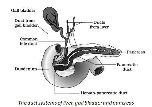
- Hepatic cells secrete bile.
- Bile is stored and concentrated in a thin muscular sac called the gall bladder.
- The duct of gall bladder is called cystic duct.
- The common hepato – pancreatic duct opens into the duodenum and opening is guarded by a sphincter called Sphincter of Oddi. Sphincter of Oddi surrounds the ampulla of Vater or Hepato pancreatic ampulla.
Functions of liver
- Secretion and synthesis of bile (daily 700-1000 ml) pH value of bile is 7.6 – 8.6
- Bile contains bile pigments (bilirubin and biliverdin), bile salts sodium glycocholate, sodium taurocholate and sodium bicarbonate.
- Carbohydrate Metabolism : Glycogenesis,Glycogenolysis, Gluconeogenesis and lipogenesis
- Deamination and formation of urea (through ornithine cycle).
- Synthesis of plasma proteins like albumins and globulins, blood clotting factors like fibrinogen and prothrombin, anticoagulants like heparin.
- Detoxification, Haemopoiesis and break down of RBC (erythroclasia)
- Lactic acid formed during anaerobic muscle contraction is converted into glycogen in the liver by Cori cycle.
- Liver acts as thermoregulatory organ
- Kupffer cells or stellat reticulo endothelial cells of liver are hepatic macrophages.
Pancreas
- It is the second largest gland
- Pancreas is a mixed gland as it has both exocrine and endocrine cells.
- Acinar cells secrete pancreatic juice.
- Pancreatic ductules arise from acini combine to form main pancreatic duct (duct of Wirsung).
- The main pancreatic duct joins the bile duct to form common hepato pancreatic duct which leads into ampulla of Vater. (hepatopancreatic ampulla) that opens into duodenum.
- Exocrine portion secretes pancreatic juice containing enzymes.
- Endocrine portion secretes hormones insulin , glucagon and other hormones
- pH value of pancreatic juice is 8.4
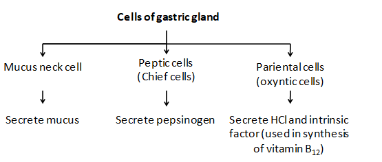
Gastric glands:
- These are present in the wall of stomach beneath the surface epithelium, these are three types
- Cardiac glands -secrete mucus for protection
- Pyloric glands – secrete mucus and the hormone gastrin (secreted by G cells).
- Fundic glands – they have three types of cells chief cells / peptic cells – secrete the proenzymes pepsinogen and prorennin neck cells – secrete mucus oxyntic cells / parietal cells – which secrete hydrochloric acid and castle’s intrinsic factor ( for absorption of vitamin B12) pH of gastric juice is 0.9 – 1.8
Intestinal glands
- They are two types – i) Brunner’s glands and ii) Crypts of Lieberkuhn
- Brunner’s glands are present in the submucosa of duodenum and secrete mucus.
- Crypts of Lieberkuhn are present in the mucosa of ileum.
- Paneth cells lining the bases of the intestinal glands secrete lysozyme (anti bacterial agent).
The process of digestion is accomplished by mechanical and chemical processes. Digestion of carbohydrates mainly takes place in mouth and small intestine. Digestion of fats is carried out in small intestine. Whereas digestion of proteins is mainly in stomach and small intestine. Finally Absorption of digested food is carried out by large intestine
4. DIGESTION PROCESS IN MOUTH
- The buccal cavity performs two major functions, mastication of food and facilitation of swallowing.
- The teeth and the tongue with the help of saliva masticate and mix up the food thoroughly.
- Mucus in saliva helps in lubricating and adhering the masticated food particles into a bolus.
Action of Saliva
- The saliva secreted into the oral cavity contains electrolytes (Na+, K+, Cl–, HCO–) and enzymes, salivary amylase and lysozyme.
- The chemical process of digestion is initiated in the oral cavity by the hydrolytic action of the carbohydrate splitting enzyme, the salivary amylase.
- About 30 per cent of starch is hydrolysed here by this enzyme (optimum pH 6.8) into a disaccharide – maltose.
- Lysozyme present in saliva acts as an antibacterial agent that prevents infections.
- The bolus is then conveyed into the pharynx and then into the oesophagus by swallowing or deglutition.
- The bolus further passes down through the oesophagus by successive waves of muscular contractions called peristalsis.
- The gastro-oesophageal sphincter controls the passage of food into the stomach.
Gastric Glands
The mucosa of stomach has gastric glands. Gastric glands have three major types of cells namely –
5. DIGESTION IN STOMACH
- The stomach stores the food for 4-5 hours. The food mixes thoroughly with the acidic gastric juice of the stomach by the churning movements of its muscular wall and is called the chyme.
- The proenzyme pepsinogen, on exposure to hydrochloric acid gets converted into the active enzyme pepsin, the proteolytic enzyme of the stomach.
- Pepsin converts proteins into proteoses and peptones (peptides).
- The mucus and bicarbonates present in the gastric juice play an important role in lubrication and protection of the mucosal epithelium from excoriation by the highly concentrated hydrochloric acid.
- HCl provides the acidic pH (pH 1.8) optimal for pepsins.
- Rennin is a proteolytic enzyme found in gastric juice of infants which helps in the digestion of milk proteins.
- Small amounts of lipases are also secreted by gastric glands.
6. DIGESTION IN SMALL INTESTINE
- Various types of movements are generated by the muscularis layer of the small intestine.
- These movements help in a thorough mixing up of the food with various secretions in the intestine and thereby facilitate digestion.
- The bile, pancreatic juice and the intestinal juice are the secretions released into the small intestine.
- Pancreatic juice and bile are released through the hepato-pancreatic duct.
- The pancreatic juice contains inactive enzymes – trypsinogen, chymotrypsinogen, procarboxypeptidases, amylases, lipases and nucleases.
- Trypsinogen is activated by an enzyme, enterokinase, secreted by the intestinal mucosa into active trypsin, which in turn activates the other enzymes in the pancreatic juice.
- The bile released into the duodenum contains bile pigments (bilirubin and bili-virdin), bile salts, cholesterol and phospholipids but no enzymes.
- Bile helps in emulsification of fats, i.e., breaking down of the fats into very small micelles. Bile also activates lipases.
- The intestinal mucosal epithelium has goblet cells which secrete mucus.
- The secretions of the brush border cells of the mucosa along with the secretions of the goblet cells constitute the intestinal juice or succus entericus.
- This juice contains a variety of enzymes like disaccharidases (e.g., maltase), dipeptidases, lipases, nucleosidases, etc.
- The mucus along with the bicarbonates from the pancreas protects the intestinal mucosa from acid as well as provide an alkaline medium (pH 7.8) for enzymatic activities.
- Sub-mucosal glands (Brunner’s glands) also help in this.
- Proteins, proteoses and peptones (partially hydrolysed proteins) in the chyme reaching the intestine are acted upon by the proteolytic enzymes of pancreatic juice as given below:
- Carbohydrates in the chime are hydrolysed by pancreatic amylase into disaccharides:
- Fats are broken down by lipases with the help of bile into di- and monoglycerides:
- Nucleases in the pancreatic juice acts on nucleic acids to form nucleotides and nucleosides:
- The enzymes in the succus entericus act on the end products of the above reactions to form the respective simple absorbable forms.
- These final steps in digestion occur very close to the mucosal epithelial cells of the intestine.
- The breakdown of bio-macromolecules mentioned above occurs in the duodenum region of the small intestine.
- The simple substances thus formed are absorbed in the jejunum and ileum regions of the small intestine.
- The undigested and unabsorbed substances are passed on to the large intestine.
- No significant digestive activity occurs in the large intestine.
- The functions of large intestine are
(i) absorption of some water, minerals and certain drugs;
(ii) secretion of mucus which helps in adhering the waste (undigested) particles together and lubricating it for an easy passage.
7. ABSORPTION
- Absorption is the process by which nutrient molecules are taken into the cells of living organisms.
- Digested food is absorbed into the blood and lymph from the alimentary canal.
- Most absorption of food occurs in the small intestine.
- Villi are short finger-like processes that project from the mucosa of small intestine having network of capillaries and lymph vessels (lacteals).
- They increase the surface area for absorption of products of digestion.
The undigested, unabsorbed substances called faeces enters into the caecum of the large intestine through ileo-caecal valve, which prevents the back flow of the faecal matter.
It is temporarily stored in the rectum till defaecation.
Absorption of Digested Products
- Absorption is the process by which the end products of digestion pass through the intestinal mucosa into the blood or lymph.
- It is carried out by passive, active or facilitated transport mechanisms.
- Small amounts of monosacharides like glucose, amino acids and some of electrolytes like chloride ions are generally absorbed by simple diffusion.
- The passage of these substances into the blood depends upon the concentration gradients.
- However, some of the substances like fructose and some amino acids are absorbed with the help of the carrier ions like Na+.
- This mechanism is called the facilitated transport.
- Transport of water depends upon the osmotic gradient.
- Active transport occurs against the concentration gradient and hence requires energy.
- Various nutrients like amino acids, monosaccharides like glucose, electrolytes like Na+ are absorbed into the blood by this mechanism.
- Fatty acids and glycerol being insoluble, cannot be absorbed into the blood.
- They are first incorporated into small droplets called micelles which move into the intestinal mucosa. They are re-formed into very small protein coated fat globules called the chylomicrons which are transported into the lymph vessels (lacteals) in the villi. These lymph vessels ultimately release the absorbed substances into the blood stream.
- Absorption of substances takes place in different parts of the alimentary canal, like mouth, stomach, small intestine and large intestine.
- However, maximum absorption occurs in the small intestine.
| Absorption in different parts of stomach | |||
| Mouth | Stomach | Small Intestine | Large intestine |
| Certain drugs coming in contact with the mucosa of mouth and lower side of the tongue are absorbed into the blood capillaries lining them. | Absorption of water, simple sugars, and alcohol etc. takes place. | Principal organ for absorption of nutrients. The digestion is completed here and the final products of digestion such as glucose, fructose, fatty acids, glycerol and amino acids are absorbed through the mucosa into the blood stream and lymph. | Absorption of water, some minerals and drugs takes place. |
- The absorbed substances finally reach the tissues which utilise them for their activities. This process is called assimilation. The digestive wastes, solidified into coherent faeces in the rectum initiate a neural reflex causing an urge or desire for its removal. The egestion of faeces to the outside through the anal opening (defaecation) is a voluntary process and is carried out by a mass peristaltic movement.
Summary of absorption in different parts of digestive system
- Mouth – Certain drugs coming in contact with the mucosa of mouth on lower surface of tongue are absorbed into blood capillaries lining them.
- Stomach – Water, simple sugars and alcohol.
- Small intestine – Principal organ for absorption of nutrients. Glucose, fructose, galactose, and amino acids are absorbed into blood. Fatty acids and glycerol are absorbed into lacteals.
- Large intestine – Water, minerals and drugs.
Assimilation
- Utilisation of the absorbed substances by the tissues is called assimilation.
Defaecation
- Digestive waste, seen as faeces in the rectum initiate a neural reflex causing an urge or desire for its removal.
- Egestion of faeces to the outside through the anal opening is a voluntary process and it is carried out by mass peristaltic movement.
- Faeces is egested to the outside through the anal
Peristalsis
- Peristalsis starts from oesophagus, continues up to rectum
- Peristalsis is slow in intestine
- Peristalsis is a part of mechanical digestion.
- Stimulation of parasympathetic nervous system results in the increase of gut peristalsis.
- Reverse peristalsis in the stomach produces vomiting.
8. DISORDERS OF DIGESTIVE SYSTEM
- Most common disorder is inflammation of the intestinal tract due to bacterial and viral infections.
- Parasites like tapeworm, roundworm, thread worm, hook worm, pin worm etc cause infections of alimentary canal.
- Jaundice – Liver is affected, skin and eyes become yellow due to deposition of bile pigments.
- Vomiting – It is the ejection of stomach contents through the mouth. A reflex action controlled by vomit centre in medulla . A feeling of nausea precedes vomiting.
- Diarrhoea – Abnormal frequency of bowel movement and increased liquidity of the faecal discharge is known as diarrhoea.
- Constipation – Faeces are retained within the rectum, as bowel movements are irregular.
- Indigestion – Food is not properly digested leading to a feeling of fullness.
Protein Energy Malnutrition (PEM)
- It includes kwashiorkor,
- If protein intake is deficient in spite of normal caloric intake it leads to kwashiorkor.
- Kwashiorkor indicated by edema enlarged liver, anorexia (lack of appetite), distended abdomen (pot belly), thin limbs, retarded growth of body and brain.
- Protein – calorie under nutrition (severe malnutrition and energy deficiency) leads to marasmus.
- Prolonged starvation results in defeacation of both protein and calorie leading to marasmus.
- Marasmus is indicated by extensive tissue and muscle wasting , dry and loose skin.
9. CALORIC VALUE
- Complete combustion of one gram of carbohydrate in bomb calorimeter in laboratory yields
4.1 k.Cal. This is called caloric value of carbohydrates. - One gram of carbohydrate yields
4.0 K Cal on the oxidation in the body , it is called physiological fuel value.
Comparison of caloric and physiological fuel value
Nutrient Caloric value Physiological fuel value
Carbohydrates 4.10 kcal 4.0 kcal
Proteins 5.65 kcal 4.0 kcal
Fats 9.45 kcal 9.0 kcal