


1. INTRODUCTION
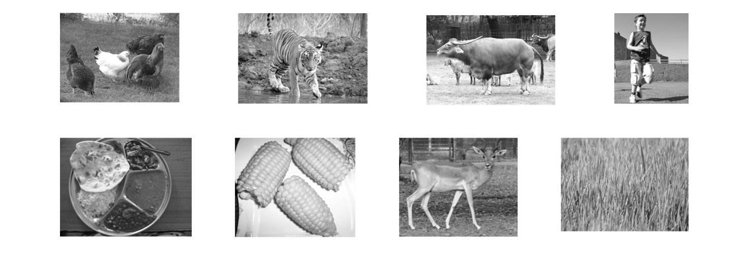
In the previous chapter, we have learnt about nutrition in plants. We know that plants photosynthesize using sunlight, water, and carbon dioxide to manufacture their own food. So, how do animals, including humans, differ from plants in the way they obtain nutrition? We have seen animals and humans eating different types of food. Look at the pictures given below. Match the animals/humans to their food.
 Thus, observe that whatever we (or animals) eat is either a plant or an animal product. But have you ever thought what happens to the food inside the body? The food substances are broken down in the animal’s body into simpler substances which can be then utilized by the body to provide energy.
2. Steps In nutrition
Whatever may be the food and the nutrients present in the food, it undergoes the following five steps: These five steps are the steps in the process of digestion.
(i) Ingestion: The process by which food is taken in by the organisms is called ingestion.
(ii) Digestion: It is the process of breaking down of complex food into simpler absorbable molecules. This is brought about with the help of special molecules called digestive juices or enzymes.
(iii) Absorption: It is the process by which digested food is taken up (or absorbed) by the body.
(iv) Assimilation: The absorbed food is incorporated into living cells and is used by the body for its growth and other purposes.
(v) Egestion: This is the process by which undigested food is removed from the body.
Nutrition: The process by which organisms obtain and use food is known as nutrition.
Enzymes: Enzymes are proteins in nature. These act as catalysts.
Nutrition in different animals
Different organisms have different ways of taking in food.
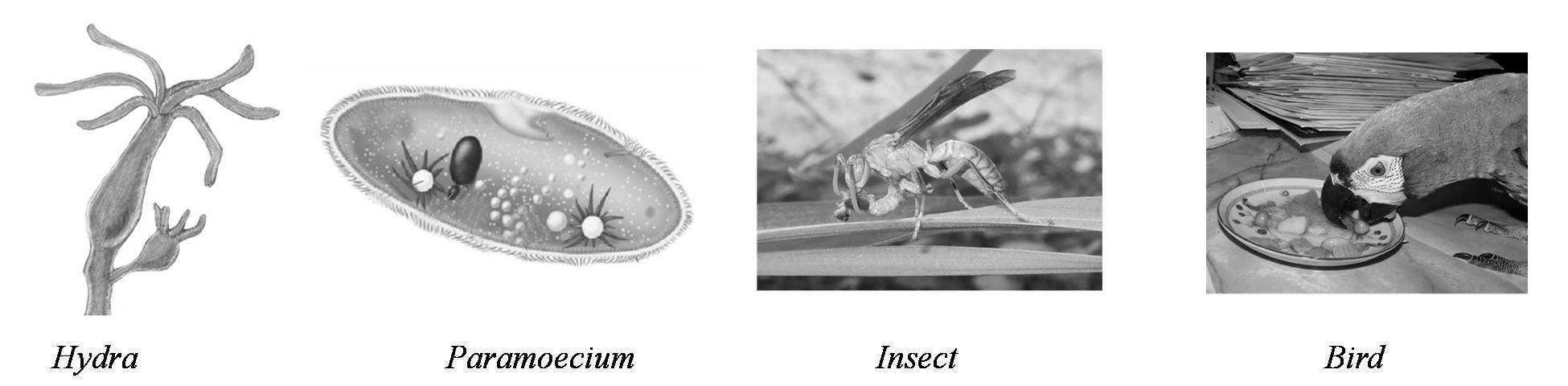
In some animals, food is taken in with the help of outgrowths on their bodies, like tentacles in Hydra and cilia in Paramecium. Small animals like insects and worms have special mouth parts for ingesting food. The mouth of a bird is modified into a beak, which helps it to catch food and tear or crack it. In this chapter we will learn about nutrition in different animals.
3. NUTRITION IN AMOEBA
i) Amoeba is an organism made up of a single cell. It is usually found in ponds and ditches.
ii) Its food consists of the microscopic plants and animals floating in these waters. When Amoeba comes in contact with its food, it throws out finger-like projections from its body.
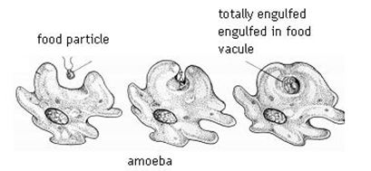
iii) These projections are called pseudopodia (pseudo, false; podium, feet). The pseudopodia completely enclose the food forming small cavities called food vacuoles.
iv) The food is digested in the food vacuole with the help of digestive juices which are secreted into the food vacuole.
v) This digested food is now ready to be absorbed and assimilated. The undigested food is pushed out of the body.
4. NUTRITION IN HUMAN BEINGS [DIGESTION IN HUMANS]
The body cells cannot use the food in the form it is eaten by us. It is converted into a simpler form by the process of digestion.
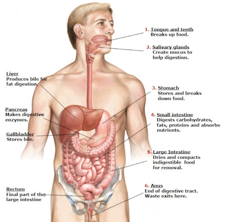
The process of digestion starts in the mouth. From the mouth, the food passes through a food canal (called alimentary canal).
Alimentary canal is a long, muscular and coiled tube. It starts from the mouth and ends at anus.
Why do we need a Digestive System?
All animals need food to survive, grow and function properly. Often the food eaten is solid. Solid food cannot be absorbed by the body cells as it is. Our digestive system has two basic jobs to do with the food we take in. The first job is to break down large food particles so that they can be carried through the body.
The second job of the digestive system is to transform the molecules of food into simple molecules. We take in foods of all kinds—milk, meat, tea, potato, fish, and so on. These molecules must be broken down into simpler molecules, so that they can finally be built into human protoplasm. Digestive system ensures that the digested material is absorbed into the blood vessels. In this way nourishment reaches all the body cells.
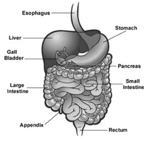
The different organs of the alimentary canal are as follows:
1. Mouth and mouth cavity
2. Oesophagus (gullet)
3. Stomach
4. Small Intestine
5. Large Intestine
6. Anus
Associated with the alimentary canal are some glands. These are:
1. Salivary glands
2. Liver
3. Pancreas.
The alimentary canal along with the associated glands is called the digestive system (Figure).
I. Mouth
The mouth contains the tongue, teeth and salivary glands. Process of digestion starts in the mouth itself. Food is bitten off and chewed (masticated) by the teeth. The chewed food gets mixed with the saliva secreted by the salivary glands. The tongue helps in mixing the food with saliva and its swallowing down the digestive system.
Teeth: There are four main kinds of teeth in man – incisors, canines, premolars and molars (Figure and Table).
The front four teeth in each jaw are the incisors. They are flat and help in biting the food. On either side of the incisors are the canines. These are sharp and two in number in each jaw. They are meant for tearing the food.
|
Different types of teeth
|
||||||||||||||||||
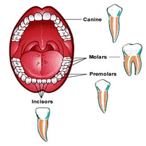
The premolars and molars are meant for grinding and crushing the food. Premolars are behind the canines, two in number on either side in each jaw. Molars are behind the premolars. In an adult, they are six in number in each jaw, three each on either side of the premolars.
In young people, there are 8 molars in all. The second set of 4 molars appears at the age of eighteen or even later. These are called the wisdom teeth.
Each jaw in an adult has 16 teeth, or 32 teeth in all.
Man has two sets of teeth – – milk teeth and permanent teeth. The first set of teeth in a baby are called milk teeth. These are replaced by permanent teeth when one is a child.
• At birth, a human infant has no teeth. After six months or so, the first teeth appear in the centre of the lower jaw.
•Milk teeth are twenty in number.
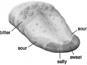
Tongue: Tongue is-: also important for eating. It helps in Bitter mixing the chewed food with saliva and swallowing the food. Further, the tongue tastes, as it has sense organs called the taste buds. These buds distinguish four basic tastes – salty, sour, sweet and bitter (Figure). In addition, tongue helps us to speak.
Salivary glands: There are three pairs of salivary glands in our mouth. A watery material called saliva is secreted by these glands. Saliva helps in the digestion of food. Saliva contains an enzyme called amylase (also called ptyalin). Amylase acts on starch and changes it into a sugar (called maltose). This sugar is sweet and soluble in water.
Note: Liver is the largest gland in the body.
II. Oesophagus (Gullet)
Oesophagus connects the mouth cavity with the stomach, and is also called the food pipe. No digestion takes place here. It only helps in pushing the food into stomach.
III. Stomach
The stomach is a muscular bag lying in the upper abdomen. Here the food is churned and converted into a semi-solid paste. The stomach secretes a juice called gastric juice and an acid. Proteins present in the food are digested by the gastric juice partly. The partly digested food from here goes to the small intestine.
IV. Small Intestine
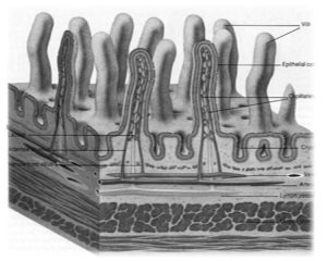
Small intestine is a long coiled tube. It also secretes a juice and digestion of all types of food is carried out here. As a result of digestion, food is converted into simple form, and glucose, amino acids and fatty acids, etc., are formed. These end products are ready for absorption. The inner surface of the small intestine has a number of finger-like projections called villi (Figure). These villi increase the area for absorption of digested food.
Saliva contains an enzyme called amylase {also called ptyalin). Amylase acts on starch and changes it into a sugar (called maltose). This sugar is sweet and soluble in water. Small intestine also absorbs the digested food and passes it on to the blood system. Thus, the nutrients are carried to all parts of the body.
Note: Small intestine is larger in length (about 6 metres) than the large intestine (about 1.5 metres). It is the main organ for the absorption of digested food.
V. Large Intestine
Large intestine has no digestive function to carry out. It helps in absorbing water and in removing the undigested solid wastes through the anus.
Liver and Pancreas: These are special organs connected with the digestive system. The liver secretes juices which help in digestion and are stored in a small bag called the gall bladder (Figure). The pancreas secretes a substance called insulin and also a juice. Insulin is important for regulating sugar level in the body.
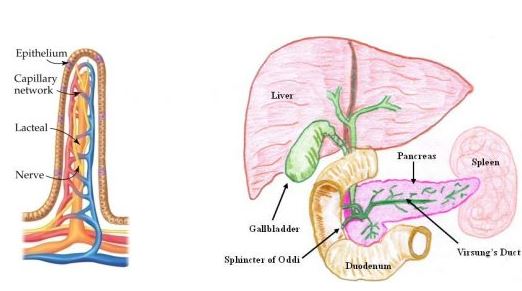
Villi in the small intestine Associated glands – liver and pancreas – with the alimentary canal
5. Digestion In Humans
We have already studied the digestive system of humans. The process of digestion is brought about with the help of digestive juices called enzymes.
i) The process of digestion starts in the mouth itself where the food is mixed with saliva. Saliva, secreted by the salivary glands, contains digestive juices which help in the breakdown of starch into sugar. The saliva also makes the food slimy so that it can be easily swallowed.
ii) The food passes from the mouth into a long tube called the oesophagus (also called food pipe).
iii) The walls of the oesophagus contracts and relaxes to produce wave-like movements (called peristaltic movements). This movement helps to move the food down into a large sac-like muscular organ called the stomach.
iv) Further digestion of food takes place in the stomach. The inner wall of the stomach secretes digestive juices, hydrochloric acid, and mucus.
v) The digestive juices help in the breakdown of proteins into simpler forms.
vi) The hydrochloric acid kills microorganisms and provides an acidic medium for effective digestion.
vii) After digestion in the stomach, the semi-digested food called the chyme passes into the small intestine.
viii) Further digestion of food takes place in the small intestine where the secretions of the liver and pancreas are released.
ix) The liver secretes bile which plays an important role in the digestion of fats. Bile is stored in an organ called gall bladder before being released in the small intestine.
x) The secretions of the pancreas called pancreatic juice help in the breakdown of carbohydrates into sugars, proteins into amino acids, and fats into fatty acids and glycerol. Thus, the digestion of various components of food is completed in the small intestine.
| SUMMARY OF DIGESTION | |||
| Part of the Digestive System | Enzymes/Juices | Food acted upon | Products formed |
| Mouth | Saliva (amylase) | Starch | Sugars |
| Stomach | Gastric juice (pepsin) | Proteins | Amino acids |
| Pancreas | Pancreatic juice (several enzymes like trypsin, lipase and sucrase) | Proteins, starch, fats | Amino acid, maltose, fatty acids, glucose |
Absorption
i) Even though digestion continues in the small intestine, the main job of the small intestine is to absorb the nutrients.
ii) The lining of the small intestine has finger-like projections called villi (singular: villus) that increase the surface area of the lining.
iii) This makes absorption more efficient. Each villus has a network of fine blood vessels.
iv) Nutrients are absorbed into the blood present in these fine blood vessels.
v) The chyme now passes into the large intestine where only water and minerals remain to be absorbed.
Assimilation
The nutrients that are absorbed in the blood are transported to the rest of the body. The final product of carbohydrate digestion, namely glucose, is broken down in the cells with the help of oxygen into carbon dioxide and water to release energy. Amino acids are used for repairing worn out cells and tissues. Fatty acids and glycerol act as energy reserve and are stored for further use.
Egestion
After absorption of water in the large intestine, the undigested food becomes semi-solid. It is then stored in the rectum until it is excreted via the anus.
Diarrhoea
Sometimes, consumption of infected food or unclean water can result in a condition called diarrhoea. It is an infection of the intestine and involves passing of watery stools very frequently. This leads to the loss of useful salts from the body and can cause dehydration. Dehydration is a condition that results due to loss of water from the body and can be serious. It can be avoided by giving Oral Rehydrating Solution (ORS) to the patient suffering from diarrhoea. The World Health Organization (WHO) has recommended a simple ORS remedy that can be made at home by simply adding salt and sugar in water.ORS sachets are also easily available in all chemist stores.
6. Teeth In Herbivores, Carnivores and Omnivores
Dentition: The arrangement of teeth in the jaws is called dentition.
Teeth in herbivores (like rabbit), carnivores (like dog) and omnivores (like human beings) are related to the diet the animals take.
As mentioned earlier, there are four different kinds of teeth
(i) incisors (for cutting) (ii) canines (for tearing)
(iii) premolars, (iv) molars (for grinding)
Dentition In Herbivores (Example Rabbit)
They have incisors which are sharp and used for cutting. Canines are absent and a gap occurs between the incisors and premolars. This gap is called diastema. The premolars and molars are used for chewing.
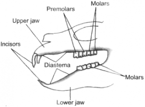
Dentition in a rabbit
The premolars and molars are almost similar in shape and size, as they have the same function. The gap between the incisors and the premolars (diasteina) allows the tongue to manipulate the food.
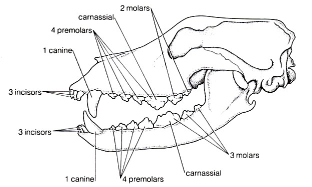
Dentition In Carnivores(Example Dog)
In carnivores the teeth in different regions of the mouth are specialised to perform a particular function. They have all four kinds of teeth. The incisors in the front of the mouth grip the food and strip off small pieces of flesh. The canines are long, sharp and pointed and adapted for flesh eating. The molars have somewhat flat surface for grinding and crushing the bones.
Dentition in omnivores (example human beings)
In omnivores, all the four kinds of teeth are well-adapted to cope up with a wide range of foods the vegetables and variety of meat. The teeth are not as specialised as in carnivores and herbivores.
The four kinds of teeth in human beings have been already described.
Structure of a Tooth
You have learnt that teeth, present in the mouth cavity, play an important role in biting and chewing the food. In an adult human being, there are four types of teeth – – incisors, canines, premolars and molars.
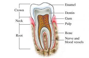
Fixed to the gums, each tooth has following three parts (Figure):
A tooth in section
(i) Root, the part embedded in the jaw,
(ii) Crown, the top part projecting above the gums, and
(iii) Neck, the part between the root and the crown.
Internally, there are three parts of a tooth – enamel, dentine and the pulp cavity. Enamel is the white part of the tooth, and is the hardest substance in our body. Below the enamel, dentine is present. Inner to dentine is the soft pulp cavity which contains blood vessels and nerves (Figure).
8.Care of The Teeth
Teeth are commonly seen to become yellow and suffer from cavity formation. When food is eaten, small amounts are left in between the teeth. Saliva and bacteria (present in the mouth) stick to the teeth and form a sticky film. This sticky film is called dental plaque.
The enzymes produced by the bacteria act on the food particles, particularly sugars and acids are produced. The acids dissolve away the tooth enamel causing tooth decay. Ultimately a hole or cavity is formed in the tooth.
Plaque affects the gums too, causing gum disease. The gums swell and may bleed on brushing.
The plaque builds up in the region where the teeth meet the gums, and forms a space. Bacteria growing in this space cause the teeth to fall.
Plaque formation and other diseases can be avoided by
(i) not eating foods like sweets, chocolates and ice-creams,
(ii) consuming foods which contain sufficient calcium, phosphorous and vitamin D such as milk, fish, raw vegetables, carrots, spinach (palak), radish, cabbage and fresh fruits (the teeth and gums get good exercise when fibrous foods are eaten),
(iii) cleaning teeth after eating sweet, sticky food,
(iv) brushing teeth thoroughly and regularly everyday in the morning and again before going to bed, and
(v) using fluoride toothpaste.
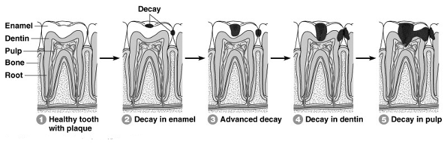
Tooth decay – stages
9. NUTRITION IN RUMINANTS
Have you seen a cow chew continuously and ever wondered why they do so?
Actually, cows and some other plant-eating animals swallow large amount of food at a time. Later, they bring back the swallowed food into the mouth to chew on it again. This procaess is called rumination and such animals are called ruminants. This habit of plant-eating animals makes their digestive system different from us and thus the need to study them separately.
I. Ingestion
The food is taken into the mouth with the help of the tongue. As the food is being chewed, it is also mixed with saliva. Since grass is difficult to digest, ruminants have big chewing teeth with powerful jaw muscles.
Note
A cow makes 40,000 to 60,000 jaw movements per day, while it keeps on chewing and rechewing.
II. Digestive system
After the initial chewing, the food passes down the 2 to 3 feet long oesophagus. The oesophagus leads into the stomach, which in ruminants is four chambered. Thus, you see that ruminants have a unique stomach as we shall soon see Thus, you see that ruminants have a unique stomach as we shall soon see.
a. Rumen
The rumen helps in storing the large quantities of food that has been quickly consumed. The food is partially digested here and is now called the Cud. The cud is then brought back into the mouth, re-chewed, and re-swallowed in a process called Cud-chewing. The rumen also has billions of bacteria and protozoa which breakdown the fibre called cellulose found in hay and grass.
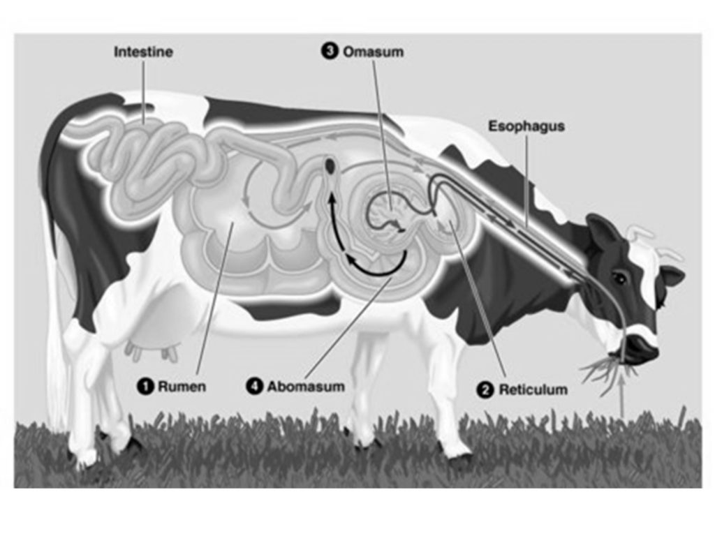
b. Recticulum
The reticulum helps in moving the swallowed food back into the mouth for thorough chewing. The reticulum opens into the Omasum.
The reticulum is informally also called the hardware stomach because if cows accidentally eat hardware (such as a piece of metal), it will often lodge here causing no further damage.
c. Omasum
The main function of Omasum is to absorb excess water and reduce the particle size further.
d. Abmosaum
The walls of the abomasum secrete digestive juices that help in digestion.
III. Absorption
Absorption begins in the four-chambered stomach itself but the main absorptive organs are the intestines.The food from the abomasum passes into the small intestine, where it mixes with secretions from the pancreas and liver. Most of the digestion of carbohydrates, proteins, and fats takes place here. Several villi are present here, which help in increasing the surface area for absorption. The small intestine leads into the large intestine. The main function of the large intestine is to absorb water from the digested food that is flowing down, make it more solid, and excrete it out as faeces.